Review
. 2011 Jul 1;3(7):a004754.
doi: 10.1101/cshperspect.a004754.

Regulation of cholesterol and fatty acid synthesis

Affiliations
Review

Regulation of cholesterol and fatty acid synthesis

Jin Ye et al. Cold Spring Harb Perspect Biol. .

Abstract

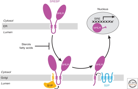
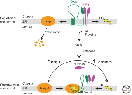
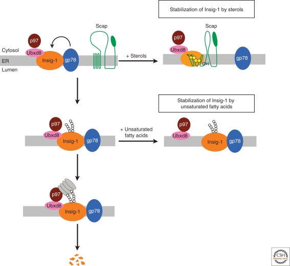
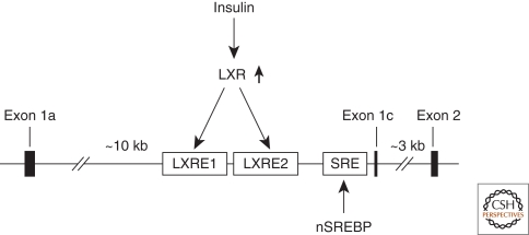
In mammals, intracellular levels of cholesterol and fatty acids are controlled through a feedback regulatory system mediated by a family of transcription factors called sterol regulatory element-binding proteins (SREBPs). SREBPs are synthesized as inactive precursors bound to membranes of the endoplasmic reticulum. When cells are deprived of cholesterol and fatty acids, NH(2)-terminal fragments of SREBPs become proteolytically released from membranes and migrate to the nucleus to activate transcription of genes required for lipid synthesis and uptake. Conversely, lipid repletion inhibits proteolytic processing of SREBPs and thereby suppresses lipid accumulation. We review here studies in cultured cells that reveal the mechanism for regulation of SREBP proteolytic activation, and those in animal models in which SREBP proteolysis has been either activated or inhibited to show the essential role of SREBPs in regulating hepatic lipid homeostasis.


Figures

Figure 1.
Figure 1.
The SREBP pathway. When cells are depleted of sterols and fatty acids, SREBPs are transported from the ER to the Golgi apparatus, in which they are first cleaved by the Golgi-localized Site-1 Protease (S1P). S1P cleaves SREBPs in the luminal loop between the two membrane-spanning sequences. Once the two halves of the SREBP are separated, a second Golgi protease, Site-2 Protease (S2P), cleaves the NH2-terminal bHLH-Zip domain of SREBPs at a site located three residues within the membrane-spanning region. After the second cleavage, the NH2-terminal domain is released from the membrane and enters the nucleus, in which it activates genes controlling lipid synthesis.
Figure 2.
Figure 2.
Cholesterol controls transport of SREBPs from the ER to Golgi complex by regulating the binding between Insig-1 and Scap. In cells depleted of cholesterol, Insig-1 is dissociated from Scap and degraded by proteasome. This allows the Scap/SREBP complex to be incorporated into COP-II-coated vesicles and transported to the Golgi complex, in which SREBPs are proteolytically activated. The NH2-terminal domain of SREBPs enters the nucleus to activate genes required for cholesterol synthesis as well as the gene encoding Insig-1. The proteolytic processing of SREBPs will not be terminated until two SREBP-induced products converge on Scap simultaneously: (1) Newly synthesized cholesterol accumulated in the ER that induces a conformational change in Scap, resulting in its increased affinity with Insig-1; and (2) Newly synthesized Insig-1 that interacts with Scap. In these cells with cholesterol restoration, binding between Scap and Insig-1 stabilizes Insig-1 and prevents incorporation of the Scap/SREBP complex into COP-II-coated vesicles.
Figure 3.
Figure 3.
Unsaturated fatty acids and sterols inhibit Insig-1 degradation independently. In sterol- and fatty acid-depleted cells, Insig-1 is ubiquitinated by gp78. Ubxd8 recruits the ATPase p97 to Insig-1. These two signals on Insig-1 lead to recognition and subsequent degradation of Insig-1 by proteasomes. Sterols induce binding of Scap to Insig-1, a reaction that displaces gp78 from Insig-1. As a result, ubiquitination of Insig-1 is inhibited. In contrast, sterols do not inhibit binding of the Ubxd8/p97 complex to Insig-1. Unsaturated fatty acids do not affect ubiquitination of Insig-1; instead, they block the interaction between Insig-1 and Ubxd8, thereby preventing the recruitment of p97 to Insig-1. Inasmuch as proteasome binding requires both ubiquitination and p97, either sterols or unsaturated fatty acids can block the degradation of Insig-1.
Figure 4.
Figure 4.
Regulation of SREBP-1c expression in the liver. The promoter of SREBP-1c contains two LXR-response elements (LXRE) and a SRE. Nuclear SREBPs bind SRE to maintain basal transcription of SREBP-1c. Insulin stimulates the activity of LXR, which binds LXRE to activate transcription of SREBP-1c.

References

    1. Adams CM, Reitz J, De Brabander JK, Feramisco JD, Li L, Brown MS, Goldstein JL 2004. Cholesterol and 25-Hydroxycholesterol inhibit activation of SREBPs by different mechanisms, both involving SCAP and insigs 2. J Biol Chem 279: 52772–52780 - PubMed
    1. Amemiya-Kudo M, Shimano H, Yoshikawa T, Yahagi N, Hasty AH, Okazaki H, Tamura Y, Shionoiri F, Iizuka Y, Ohashi K, et al. 2000. Promoter analysis of the mouse sterol regulatory element-binding protein-1c gene. J Biol Chem 275: 31078–31085 - PubMed
    1. Brown MS, Goldstein JL 1997. The SREBP pathway: Regulation of cholesterol metabolism by proteolysis of a membrane-bound transcription factor. Cell 89: 331–340 - PubMed
    1. Brown MS, Goldstein JL 2009. Cholesterol feedback: From Schoenheimer’s bottle to Scap’s MELADL. J Lipid Res 50: S15–27 - PMC - PubMed
    1. Brown MS, Ye J, Rawson RB, Goldstein JL 2000. Regulated intramembrane proteolysis: A control mechanism conserved from bacteria to humans. Cell 100: 391–398 - PubMed

Publication types

MeSH terms

Substances