Review
. 2011 May 1;3(5):a005322.
doi: 10.1101/cshperspect.a005322.

Golgi positioning

Affiliations
Review

Golgi positioning

Smita Yadav et al. Cold Spring Harb Perspect Biol. .

Abstract

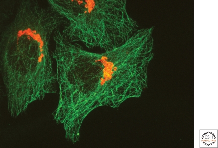
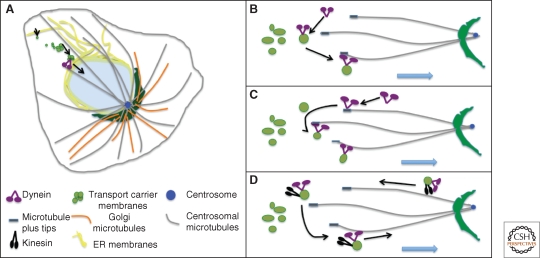
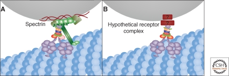
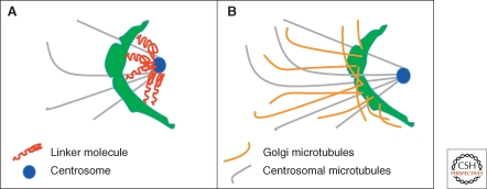
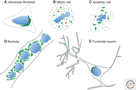
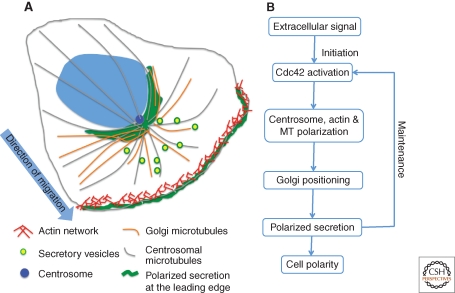
The Golgi apparatus in mammalian cells is positioned near the centrosome-based microtubule-organizing center (Fig. 1). Secretory cargo moves inward in membrane carriers for delivery to Golgi membranes in which it is processed and packaged for transport outward to the plasma membrane. Cytoplasmic dynein motor proteins (herein termed dynein) primarily mediate inward cargo carrier movement and Golgi positioning. These motors move along microtubules toward microtubule minus-ends embedded in centrosomes. Centripetal motility is controlled by a host of regulators whose precise functions remain to be determined. Significantly, a specific Golgi receptor for dynein has not been identified. This has impaired progress toward elucidation of membrane-motor-microtubule attachment in the periphery and, after inward movement, recycling of the motor for another round. Pericentrosomal positioning of the Golgi apparatus is dynamic. It is regulated during critical cellular processes such as mitosis, differentiation, cell polarization, and cell migration. Positioning is also important as it aligns the Golgi along an axis of cell polarity. In certain cell types, this promotes secretion directed to the proximal plasma membrane domain thereby maintaining specializations critical for diverse processes including wound healing, immunological synapse formation, and axon determination.


Figures

Figure 1.
Figure 1.
The mammalian pericentrosomal Golgi ribbon. Fluorescent micrograph of cultured HeLa cells stained using antibodies against tubulin (green) and the Golgi marker protein giantin (red) shows relative positions of extensive microtubule network and the Golgi membrane network.
Figure 2.
Figure 2.
Dynein and dynactin schematic diagrams. The dynein motor protein complex is assembled on two catalytic heavy chains each containing a microtubule-binding domain, a motor domain consisting of six AAA ATPase modules, and a coiled-coil linker region (A). The linker region binds to intermediate chains and light intermediate chains and the assembled intermediate chains bind light chains. The multisubunit dynactin complex has a projecting arm comprised of dimeric p150 that binds dynein intermediate chains and microtubules and a rod-shaped core comprised of an Arp1 filament that can bind spectrin on membranes (B).
Figure 3.
Figure 3.
Inward membrane movement. A pericentrosomal Golgi ribbon network is depicted (A). Minus-ends of microtubules converge at the centrosome and support inward movement by dynein of secretory and Golgi membranes derived from the ER. Also shown are three modes of initiating membrane motility. The motor can be recruited directly from the cytosol onto membranes, which then load onto a microtubule (B). The motor can be preloaded on microtubule plus tips, which then probe the cytoplasm and capture membranes (C). The motor can be carried to the cell periphery via recycling vesicles bearing active kinesin and then, on fusion with ERGIC membranes, becomes activated for inward motility (D).
Figure 4.
Figure 4.
Membrane recruitment of dynein. Golgi membranes are moved inward by dynein moving on microtubules and one possibility is that dynactin links dynein to Golgi membranes by binding spectrin (A). In light of dynactin-independent dynein membrane association and the nonspecific localization of spectrin, another possibility is a yet-to-be-identified receptor complex for dynein that is Golgi-specific (B).
Figure 5.
Figure 5.
Additional Golgi positioning mechanisms. Golgi membranes may be anchored directly to the centrosome and/or minus-ends of microtubules (A). Golgi microtubules contribute to Golgi structure and maintenance of Golgi positioning once the membranes have moved inward (B).
Figure 6.
Figure 6.
Dynamic changes in Golgi positioning. Golgi membranes are positioned adjacent the centrosome in many mammalian cell types during interphase (A) and fragment and disperse during mitosis (B) and apoptosis (C). The pericentrosomal Golgi ribbon in myoblasts is converted to dispersed Golgi ministacks in myotubes (D). Pyramidal neurons have both a somatic Golgi and dispersed Golgi-outposts, which localize to dendritic branchpoints (E).
Figure 7.
Figure 7.
Golgi positioning directs secretion to cell leading edge. Extracellular signals, such as wounding, trigger actin assembly and reorientation of the centrosome, the Golgi apparatus, and the microtubule array (A). As a consequence, secretion is directed to the leading edge. Once initiated, maintenance of polarity requires directed secretion toward the leading edge by the pericentrosomally positioned Golgi apparatus (B).

References

    1. Addinall SG, Mayr PS, Doyle S, Sheehan JK, Woodman PG, Allan VJ 2001. Phosphorylation by cdc2-CyclinB1 kinase releases cytoplasmic dynein from membranes. J Biol Chem 276: 15939–15944 - PubMed
    1. Allan VJ, Vale RD 1991. Cell cycle control of microtubule-based membrane transport and tubule formation in vitro. J Cell Biol 113: 347–359 - PMC - PubMed
    1. Arasaki K, Taniguchi M, Tani K, Tagaya M 2006. RINT-1 regulates the localization and entry of ZW10 to the syntaxin 18 complex. Mol Biol Cell 17: 2780–2788 - PMC - PubMed
    1. Askham JM, Vaughan KT, Goodson HV, Morrison EE 2002. Evidence that an interaction between EB1 and p150(Glued) is required for the formation and maintenance of a radial microtubule array anchored at the centrosome. Mol Biol Cell 13: 3627–3645 - PMC - PubMed
    1. Barcia C, Sanderson NS, Barrett RJ, Wawrowsky K, Kroeger KM, Puntel M, Liu C, Castro MG, Lowenstein PR 2008. T cells’ immunological synapses induce polarization of brain astrocytes in vivo and in vitro: A novel astrocyte response mechanism to cellular injury. PLoS One 3: e2977. - PMC - PubMed

MeSH terms

Substances

LinkOut - more resources