. 2011 Jun 24;286(25):22546-57.
doi: 10.1074/jbc.M110.217703. Epub 2011 Apr 19.

Embryonic toxin expression in the cone snail Conus victoriae: primed to kill or divergent function?

Affiliations

Embryonic toxin expression in the cone snail Conus victoriae: primed to kill or divergent function?

Helena Safavi-Hemami et al. J Biol Chem. .

Abstract

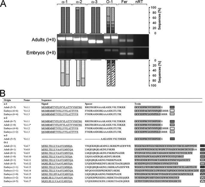
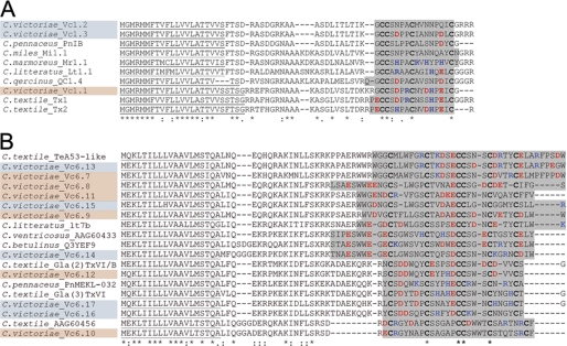
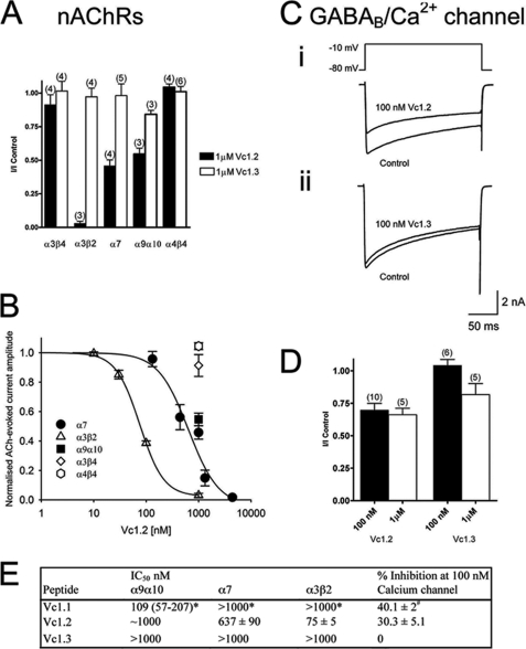
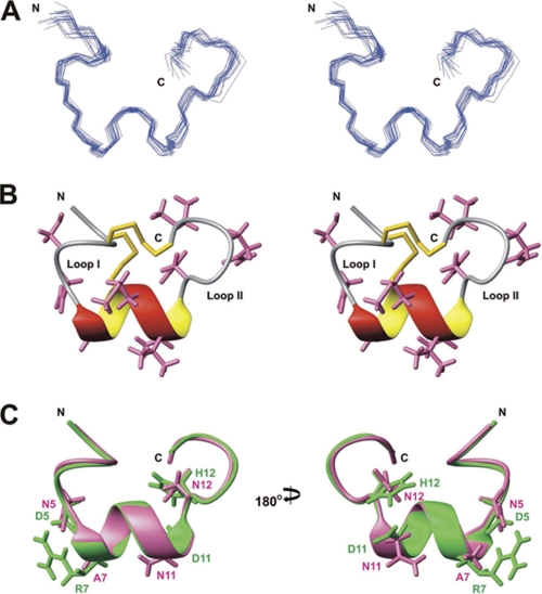
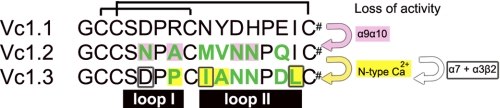
Predatory marine cone snails (genus Conus) utilize complex venoms mainly composed of small peptide toxins that target voltage- and ligand-gated ion channels in their prey. Although the venoms of a number of cone snail species have been intensively profiled and functionally characterized, nothing is known about the initiation of venom expression at an early developmental stage. Here, we report on the expression of venom mRNA in embryos of Conus victoriae and the identification of novel α- and O-conotoxin sequences. Embryonic toxin mRNA expression is initiated well before differentiation of the venom gland, the organ of venom biosynthesis. Structural and functional studies revealed that the embryonic α-conotoxins exhibit the same basic three-dimensional structure as the most abundant adult toxin but significantly differ in their neurological targets. Based on these findings, we postulate that the venom repertoire of cone snails undergoes ontogenetic changes most likely reflecting differences in the biotic interactions of these animals with their prey, predators, or competitors. To our knowledge, this is the first study to show toxin mRNA transcripts in embryos, a finding that extends our understanding of the early onset of venom expression in animals and may suggest alternative functions of peptide toxins during development.


Figures

FIGURE 1.
FIGURE 1.
A, universal α- and O-conotoxin RT-PCR showing toxin mRNA expression in embryos (n = 2, each pooled from ∼50 embryos) and adult specimens (n = 2) of C. victoriae. 110 ng of cDNA was amplified using three α- (α-1, α-2, and α-3) and one O-conotoxin (O-1) oligonucleotide pair. Ferritin (Fer) served as a reference gene. No reverse transcriptase controls (nRT) were performed to rule out contamination by genomic DNA. RT-PCRs were subsequently cloned and subjected to nucleotide sequencing. Bars represent the number of clones identified per RT-PCR expressed as percentages. DNA ladder: 1 kb plus (Invitrogen). *, analogue of Vc1.1 carrying one amino acid substitution. B, conotoxin sequences identified by RT-PCR showing the predicted signal peptide sequences (underlined), the spacer region, and the mature toxin region highlighted in gray. The number of clones sequenced per replicate (n = 2) is given in parentheses (43). GenBankTM accession numbers are as follows: Vc1.2, GU046308; Vc1.3, GU046309; Vc6.7, JF433900; Vc6.8, JF433901; Vc6.9, JF433902; Vc6.10, JF433903; Vc6.11, JF433904; Vc6.12, JF433905; Vc6.13, JF433906; Vc6.14, JF433907; Vc6.15, JF433908; Vc6.16, JF433909; and Vc6.1, JF433910.
FIGURE 2.
FIGURE 2.
Comparative alignment of novel embryonic α- (A) and O-conotoxins (B) isolated from C. victoriae with toxin sequences from other cone snail species. Alignment was performed using MAFFT E-INS-i sequence alignment by means of local pairwise alignment information (28). Sequences obtained from C. victoriae embryos and adults are highlighted blue and orange, respectively. Predicted protein signal sequences are underlined (SignalP). Conserved cysteine residues are shown in boldface, and the predicted mature toxin regions are highlighted gray (50). Basic and acidic amino acids are shown in blue and red, respectively. Dashes denote gaps. Amino acid conservations are denoted by an asterisk, and colons and periods represent a high and low degree of similarity, respectively.
FIGURE 3.
FIGURE 3.
Vc1.2 and Vc1. 3 inhibition of nAChRs expressed in Xenopus oocytes and Ca2+ channel currents in rat DRG neurons. A, bar graph of the relative inhibition of nAChRs expressed in Xenopus oocytes by 1 μm Vc1.2 and Vc1.3. Vc1.2 (1 μm) completely inhibited α3β2 nAChRs and inhibited α7 and α9α10 by 54 and 45%, respectively. Vc1.3 was inactive at all neuronal nAChR subtypes. B, concentration-response curves obtained for Vc1.2 inhibition of nAChR subtypes. Vc1.2 was most active at α3β2 nAChRs with an IC50 of 75 ± 5 nm (▴; n = 3), 637 ± 90 nm for α7 (●; n = 4–6), and ∼1 μm for α9α10 (■; n = 3). C, superimposed depolarization-activated whole-cell Ba2+ currents elicited by voltage steps from a holding potential of −80 to −10 mV in the absence (control) and presence of 100 nm Vc1.2 (panel i) and 100 nm Vc1.3 (panel ii), respectively. D, bar graph of the relative inhibition of HVA Ca2+ channel currents in rat DRG neurons by 100 nm and 1 μm Vc1.2 and Vc1.3. Numbers in parentheses reflect number of cells. E, comparison of the IC50 values of Vc1.1, Vc1.2, and Vc1.3 at different nAChR subtypes and percentage inhibition of calcium currents in isolated DRG neurons. Values determined in this study represent mean ± S.E. * indicates mean plus 95% confidence interval (54), and # represents mean ± S.E. (24).
FIGURE 4.
FIGURE 4.
Solution structure of Vc1.2. A, stereo view of the family of 20 final structures of Vc1.2, superimposed over the backbone heavy atoms. B, stereo ribbon view of the closest-to-average structure of Vc1.2 highlighting the α-helix and two disulfide bonds. Side chains are shown except for prolines. C, comparison of the solution structures of Vc1.2 (pink) and Vc1.1 (30) (Protein Data Bank code 2H8S, green). Structures are superimposed over the backbone heavy atoms. Side chains of Vc1.1 residues important for α9α10 nAChR binding (30) and equivalent residues in Vc1.2 are shown.
FIGURE 5.
FIGURE 5.
Alignment of α-conotoxins from C. victoriae highlighting residues important for biological function. Differences in sequence between the embryonic toxins Vc1.2 and Vc1.3 to the adult toxin Vc1.1 are shown in green. Amino acid residues likely to have caused a loss in activity toward the α9α10 neuronal nicotinic subtype (54), the N-type calcium channels,3 and the α7 subtype (55) are highlighted in pink and yellow and depicted with a frame, respectively. The disulfide bonds between Cys1–Cys3 and Cys2–Cys4 are indicated by black lines. The backbone loops formed by this disulfide connectivity are shown below the sequences. All three C termini are likely to be amidated (#), a common modification in conotoxins.
FIGURE 6.
FIGURE 6.
Histological preparations of the adult venom apparatus and embryos of C. victoriae. Sections were Mallory-stained and cut at 7 μm thickness. A, cross-section through the venom apparatus of C. victoriae showing the venom duct (VD), the venom bulb (VB), the proboscis (PB), the gill (GL), and the buccal mass (BM). Scale bar, 200 μm. B, serial sections (panels i–vi) through an embryo depicting foregut (fg), left cerebral ganglion (lcg), left pedal ganglion (lpg), left statocyst (ls), mouth (m), mantle cavity (mc), midgut (mg), midgut opening (mgo), muscle (mu), osphradial ganglia (og), osphradium (op), right cerebral ganglion (rcg), right cephalic tent (rct), right eye (re), radula sack (rs), right pedal ganglion (rpg), style sac (ss). The incipient venom gland is marked with an asterisk. Scale bar, 200 μm. C, schematic of the venom apparatus showing orientation of section shown in A. D, drawing of the larvae showing orientation of serial sections shown in B.
FIGURE 7.
FIGURE 7.
Potential usage of toxins in cone snail embryos. A, C. victoriae guarding egg capsules. Cone snail embryos may express toxin mRNA transcripts as preparation for a predatory life style (B) or to deter predators (C), or toxin peptides may be involved in other biological pathways such as neuronal signaling (D).

Similar articles

Cited by

References

    1. Terlau H., Olivera B. M. (2004) Physiol. Rev. 84, 41–68 - PubMed
    1. Olivera B. M. (2006) J. Biol. Chem. 281, 31173–31177 - PubMed
    1. Norton R. S., Olivera B. M. (2006) Toxicon 48, 780–798 - PubMed
    1. Pisarewicz K., Mora D., Pflueger F. C., Fields G. B., Marí F. (2005) J. Am. Chem. Soc. 127, 6207–6215 - PubMed
    1. Lopez-Vera E., Walewska A., Skalicky J. J., Olivera B. M., Bulaj G. (2008) Biochemistry 47, 1741–1751 - PubMed

Publication types

MeSH terms