. 2011 Jul;4(4):484-95.
doi: 10.1242/dmm.006593. Epub 2011 Apr 18.

The tumor suppressor gene Trp53 protects the mouse lens against posterior subcapsular cataracts and the BMP receptor Acvr1 acts as a tumor suppressor in the lens

Affiliations

The tumor suppressor gene Trp53 protects the mouse lens against posterior subcapsular cataracts and the BMP receptor Acvr1 acts as a tumor suppressor in the lens

Luke A Wiley et al. Dis Model Mech. 2011 Jul.

Abstract

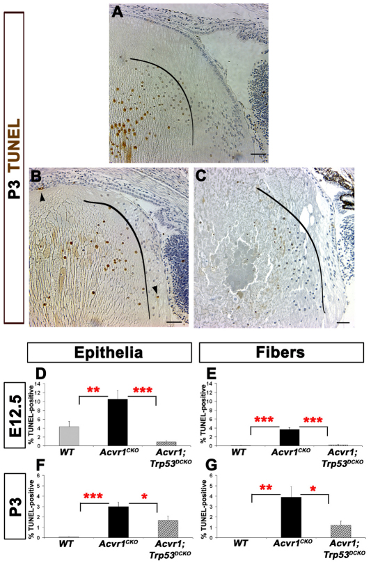
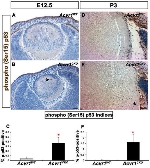
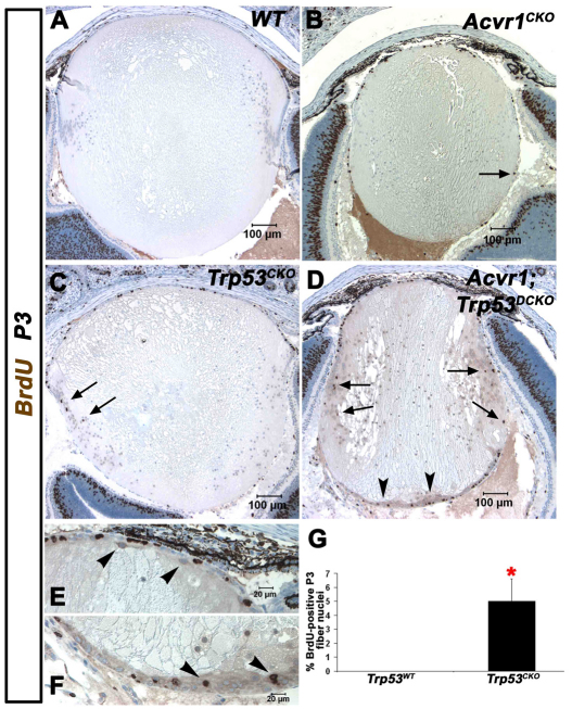
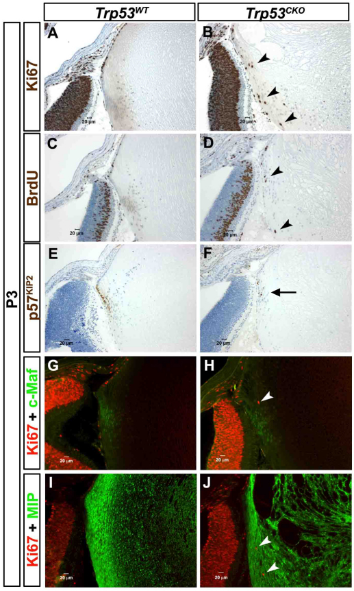
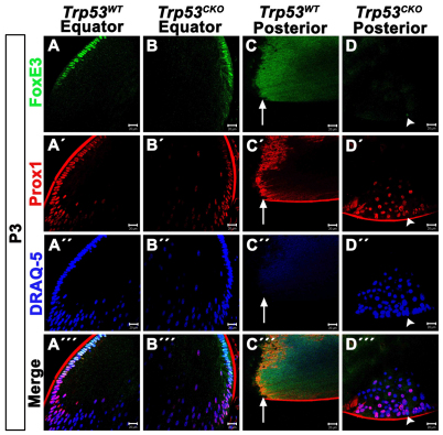
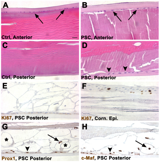
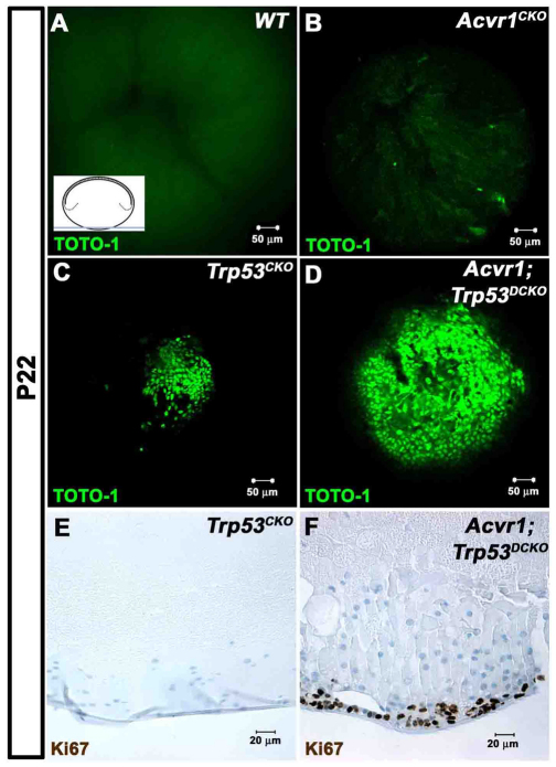
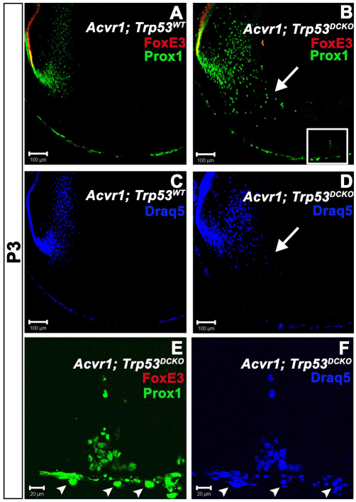
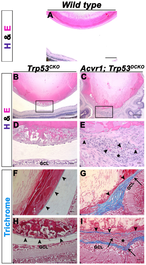
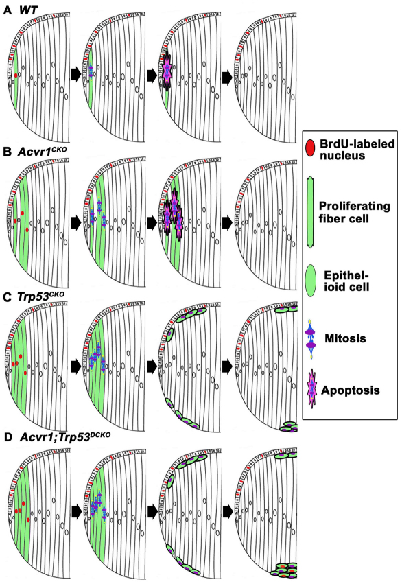
We previously found that lenses lacking the Acvr1 gene, which encodes a bone morphogenetic protein (BMP) receptor, had abnormal proliferation and cell death in epithelial and cortical fiber cells. We tested whether the tumor suppressor protein p53 (encoded by Trp53) affected this phenotype. Acvr1 conditional knockout (Acvr1(CKO)) mouse fiber cells had increased numbers of nuclei that stained for p53 phosphorylated on serine 15, an indicator of p53 stabilization and activation. Deletion of Trp53 rescued the Acvr1(CKO) cell death phenotype in embryos and reduced Acvr1-dependent apoptosis in postnatal lenses. However, deletion of Trp53 alone increased the number of fiber cells that failed to withdraw from the cell cycle. Trp53(CKO) and Acvr1;Trp53(DCKO) (double conditional knockout), but not Acvr1(CKO), lenses developed abnormal collections of cells at the posterior of the lens that resembled posterior subcapsular cataracts. Cells from human posterior subcapsular cataracts had morphological and molecular characteristics similar to the cells at the posterior of mouse lenses lacking Trp53. In Trp53(CKO) lenses, cells in the posterior plaques did not proliferate but, in Acvr1;Trp53(DCKO) lenses, many cells in the posterior plaques continued to proliferate, eventually forming vascularized tumor-like masses at the posterior of the lens. We conclude that p53 protects the lens against posterior subcapsular cataract formation by suppressing the proliferation of fiber cells and promoting the death of any fiber cells that enter the cell cycle. Acvr1 acts as a tumor suppressor in the lens. Enhancing p53 function in the lens could contribute to the prevention of steroid- and radiation-induced posterior subcapsular cataracts.


Figures

Fig. 1.
Fig. 1.
Increased apoptosis in the absence of Acvr1 is p53 dependent. The TUNEL labeling index was determined in lens epithelial and fiber cells of wild-type, Acvr1CKO and Acvr1;Trp53DCKO lenses. (A) A representative image of a TUNEL-stained Acvr1WT lens at P3. Central fibers normally denucleate, serving as a positive control for the TUNEL staining. The curved black line indicates the boundary between elongating fiber cells and deeper fiber cells undergoing the normal process of denucleation. TUNEL-positive, peripheral cortical fiber cells were rarely detected in P3 lenses. (B) Representative image of an Acvr1CKO lens at P3 showing a TUNEL-positive cortical fiber cell nucleus (upper arrowhead) and a TUNEL-positive epithelial cell (lower arrowhead). As in our previous study (Rajagopal et al., 2008), only elliptical fiber cell nuclei were counted to determine the number of TUNEL-positive cells. Rounded nuclei inside the curved line are deeper in the lens and are undergoing the normal denucleation process. Conditional deletion of Acvr1 significantly increased the TUNEL index of epithelial cells at E12.5. (C) Representative image of an Acvr1;Trp53DCKO double knockout lens at P3. TUNEL-positive mature fiber cells undergoing the normal process of denucleation are to the left of the curved line. In Acvr1;Trp53DCKO lenses, TUNEL-positive superficial fiber cells were rarely detected. (D,E) Deletion of Trp53 reduced the Acvr1-induced apoptosis in E12.5 epithelial (D) and fiber (E) cells. (F,G) A significantly lower percentage of TUNEL-positive epithelial (F) and fiber (G) cells was detected at P3 when Acvr1 and Trp53 were deleted together, compared WITH Acvr1 alone. *P<0.05; **P<0.01; ***P<0.001. Scale bars: 50 μm.
Fig. 2.
Fig. 2.
Acvr1CKO lenses have increased phosphorylation of p53 at Ser15. Acvr1WT and Acvr1CKO lenses were stained for phosphorylated (p)-p53. (A) A representative image of an E12.5 Acvr1WT lens with no detectable p-p53 staining. (B) p-p53-positive fiber cell nuclei were detected in Acvr1CKO lenses at E12.5 (arrowhead). (C) The percentage of p-p53-positive fiber cells was significantly higher in Acvr1CKO lenses than in Acvr1WT lenses at E12.5. (D) A representative Acvr1WT lens with no detectable p-p53-positive cells at P3. (E) An Acvr1CKO lens with a p-p53-positive fiber cell nucleus at P3 (arrowhead). (F) At P3, significantly more p-p53-positive fiber cells were detected in Acvr1CKO lenses than in Acvr1WT lenses. *P<0.05. Scale bars: 20 μm (A,B), 50 μm (D,E).
Fig. 3.
Fig. 3.
Acvr1;Trp53DCKO lenses develop posterior subcapsular plaques of actively proliferating cells. Lenses were injected with BrdU at P3 to label cells in S-phase of the cell cycle. (A) The normal morphology of wild-type lenses. BrdU incorporation is restricted to the anterior epithelium. (B) Acvr1CKO lenses are smaller and have cortical fiber cells that incorporate BrdU (arrow). (C) Trp53CKO lenses have aberrant fiber cells that incorporate BrdU (arrows). (D) Acvr1;Trp53DCKO lenses have numerous BrdU-positive fiber cell nuclei (arrows), more than either Acvr1CKO or Trp53CKO lenses, and form posterior subcapsular plaques of epithelioid cells, some of which are BrdU positive (arrowheads). (E) Acvr1;Trp53DCKO lenses have ectopic, spindle-shaped cells beneath the anterior epithelium (arrowheads). (F) Ectopic cells at the posterior of Acvr1;Trp53DCKO lenses (arrowheads). (G) The percentage of BrdU-positive cortical fiber cell nuclei was significantly higher in Trp53CKO lenses than in Trp53WT lenses. *P<0.05.
Fig. 4.
Fig. 4.
A subset of Trp53CKO fiber cells fail to exit the cell cycle, but express fiber-cell-specific markers. P3 lenses were stained with antibodies that label markers of cell proliferation, cell cycle exit and lens fiber cell differentiation. (A) Labeling for Ki67, a marker for actively cycling cells, is restricted to the anterior epithelium of Trp53WT lenses. (B) In Trp53CKO lenses, a few cortical fiber cells are Ki67 positive (arrowheads). (C) BrdU is only incorporated into anterior epithelial cells in Trp53WT lenses. (D) A few Trp53CKO fiber cells are BrdU positive (arrowheads). (E) p57KIP2, a marker of cell cycle exit, is strongly expressed in all nuclei in the lens equator of Trp53WT lenses. (F) Most cells in Trp53CKO lenses have nuclei that are stained with antibody to p57KIP2, but a few nuclei are p57KIP2 negative (arrow). (G) Ki67 labeling is lost near the equator, where the fiber-specific transcription factor, Maf (c-Maf) is first expressed. (H) Fiber cells in Trp53CKO lenses express Maf, although some are also Ki67 positive (arrowhead). (I) MIP, a major component of fiber cell membranes, is strongly expressed throughout the fiber zone of Trp53WT lenses. (J) MIP is strongly expressed in Trp53CKO fiber cells, even when they express Ki67 (arrowheads).
Fig. 5.
Fig. 5.
Epithelioid cells that constitute the posterior plaques in Trp53CKO lenses express markers of fiber cell terminal differentiation. Trp53WT and Trp53CKO P3 lenses were double stained with FoxE3 and Prox1. (A) FoxE3 is expressed in anterior epithelial cells, but is lost near the lens equator in Trp53WT lenses. (A′) Prox1 is first expressed near the lens equator and is strongly expressed in all fiber cell nuclei in Trp53WT lenses. (A″) The dye DRAQ-5 stains all lens nuclei and allows for visualization of the lens ‘bow’ region. (A′″) Merged image of a Trp53WT lens. (B) As in Trp53WT lenses, FoxE3 labels anterior epithelial cells in Trp53CKO lenses. (B′) Prox1 is normally expressed in differentiating lens fiber cells in Trp53CKO lenses. (B″) DRAQ-5 shows the abnormal appearance of the ‘bow’ region in a Trp53CKO lens. (B′″) Merged image of a Trp53CKO lens. (C) Image of the posterior pole of the same Trp53WT lens as in A. The image is taken at the suture and the laser intensity is purposefully increased to show the fiber zone and suture. No FoxE3-positive nuclei are detected. (C′) Similarly, no Prox1-positive nuclei are present at the posterior pole; non-specific staining is present along the sutures. The anti-mouse secondary antibody used to detect Prox1 non-specifically labels the lens capsule. (C″) Staining with DRAQ-5 shows that there are no cell nuclei at the posterior pole of the Trp53WT lens. (C′″) Merged image of the posterior pole of the Trp53WT lens. Arrows in C-C′″ indicate the posterior lens suture. (D) The posterior pole of the same Trp53CKO lens as in B has a posterior subcapsular plaque. Most epithelioid cells within this plaque do not express FoxE3; a lone cell displays weak FoxE3 labeling (arrowhead). (D′) By contrast, all cells that make up epithelioid plaques express Prox1, although some cells show weak expression. The cell that is FoxE3 positive is also Prox1 positive (arrowhead). (D″) Labeling with DRAQ-5 shows the posterior subcapsular plaque in a Trp53CKO lens. Arrowhead indicates the location of the lone FoxE3-positive cell detected in this section. (D′″) Merged image of the posterior subcapsular plaque in a Trp53CKO lens. Arrowhead indicates the location of the lone FoxE3-positive cell detected in this section. Scale bars: 20 μm.
Fig. 6.
Fig. 6.
The appearance of cells in a human PSC is similar to those in Trp53CKO mouse lenses. (A) The anterior surface of a normal adult human lens is bounded by the thick anterior capsule and a thin layer of lens epithelial cells (arrows). Beneath the epithelium are layers of well-organized superficial fiber cells. (B) The anterior surface of an adult human lens with a PSC appears similar to the normal lens in A. (C) The posterior surface of the normal lens has a thinner capsule than does the anterior of the lens. Beneath the capsule are layers of superficial fiber cells. (D) The posterior of the lens with a PSC has a layer of globular ‘balloon’ cells between the capsule and the well-organized, deeper fiber cells. A layer of epithelioid cells is present on the inner surface of the capsule (arrowheads). (E) No Ki67-labeled nuclei were detected in balloon or epithelioid cells at the posterior of the lens that had PSCs. The epitope unmasking method used in the staining procedure extracted the cytoplasm from the ‘balloon’ cells, causing them to appear as ‘ghosts’. No Ki67-positive nuclei were detected in the anterior epithelium from this lens (not shown). (F) Several nuclei in the corneal epithelium from the eye with a PSC were Ki67 positive, indicating that the staining procedure worked as expected. (G) The nuclei of balloon (arrow) and epithelioid (arrowhead) cells in the PSC stained with an antibody to the fiber-cell-specific marker Prox1. Some of the empty ‘ghosts’ resulting from the epitope unmasking method are labeled with an asterisk. (H) The nuclei of balloon (arrow) and epithelioid (arrowhead) cells in the PSC stained with an antibody to the fiber-cell-specific marker Maf.
Fig. 7.
Fig. 7.
Loss of Acvr1 leads to proliferation in posterior subcapsular plaques, forming ‘lens tumors’. P22 lenses were stained with the nucleic acid stain TOTO-1 and their posterior poles were examined using a confocal microscope. The inset in A shows the plane of section and region of the lens that was viewed. (A) No nuclei were seen in the posterior poles of Acvr1WT lenses. Staining of RNA in the fiber cell cytoplasm shows the posterior lens sutures. (B) A few scattered nuclei were evident near the posterior surface of Acvr1CKO lenses. (C) A small accumulation of nuclei was present at the posterior pole of Trp53CKO lenses. (D) A large mass of cells was present at the posterior poles of Acvr1;Trp53DCKO lenses. (E) Antibody to Ki67 showed that cells in the posterior subcapsular plaques of Trp53CKO lenses were not in the cell cycle. (F) Antibody to Ki67 showed that cells in the posterior subcapsular plaques of Acvr1;Trp53DCKO lenses were still in the cell cycle.
Fig. 8.
Fig. 8.
Cells in the posterior subcapsular plaques in Acvr1;Trp53DCKO lenses express the fiber-specific transcription factor Prox1. (A) In wild-type lenses at P3, the epithelial-specific transcription factor FoxE3 labels the nuclei of all anterior epithelial cells, but was lost in the transition zone when epithelial cells began to differentiate into fiber cells. The fiber-specific transcription factor Prox1 was first expressed in the transition zone and was detected in all differentiated fiber cells that retained nuclei. (B) FoxE3 and Prox1 had normal expression patterns in Acvr1;Trp53DCKO lenses. There was an overlap in the expression patterns for FoxE3 and Prox1 in the transition zone (yellow) in both Acvr1;Trp53WT (A) and Acvr1;Trp53DCKO (B) lenses. However, the bow region in Acvr1;Trp53DCKO lenses was disorganized, with Prox1-positive nuclei scattered in the posterior of the lens (arrow). Boxed region is shown in E and F. (C) Staining Acvr1;Trp53 lenses with DRAQ-5. (D) Staining Acvr1;Trp53DCKO lenses with DRAQ-5. Arrow indicates the disorganized bow region. (E,F) A projection of z-stacks from the boxed region in B. Cells making up the posterior subcapsular plaques in Acvr1;Trp53DCKO lenses at P4 were Prox1 positive and FoxE3 negative. The nuclei of cells of the tunica vasculosa lentis (TVL) were also stained for Prox1 (arrowheads). These could be distinguished from fiber cell nuclei because they lay on the opposite side of the lens capsule.
Fig. 9.
Fig. 9.
Adult Acvr1;Trp53DCKO lenses form vascularized, tumor-like structures at their posterior poles. 4-month-old Trp53CKO and Acvr1;Trp53DCKO lenses were sectioned and stained with hematoxylin and eosin (A–E) or Masson trichrome (F–I). (A) The posterior region of a wild-type lens and the adjacent retina. (B) Overview of the posterior half of a Trp53CKO lens. (C) Overview of the posterior of an Acvr1;Trp53DCKO lens. (D) Magnified image of the boxed area in B, showing a large posterior subcapsular-like plaque and normal separation between the lens and the ganglion cell layer (GCL) of the retina. (E) Magnified image of the boxed area in C, showing a vascularized (arrowheads) tumor-like structure (asterisk) at the posterior pole of the Acvr1;Trp53DCKO lens. (F) Section from the same Trp53CKO lens in B stained with Masson trichrome, which stains the lens capsule blue (arrowheads). (G) Section from the same Acvr1;Trp53DCKO lens in C. The original lens capsule is stained blue (arrowheads). However, towards the posterior of the lens the capsule seems to splay (arrow), forming a multilayered connective tissue. (H) The posterior capsule of the Trp53CKO lens was thin, but remained intact, despite the presence of the posterior subcapsular plaque. Arrowheads indicate the location of the posterior lens capsule. (I) In the posterior of the Acvr1;Trp53DCKO lens, the original lens capsule (arrowheads) was bordered by a multilayered connective tissue (arrows). The outer surface of this connective tissue separated the tumor-like growth (asterisk) from the GCL of the retina, encapsulating the ‘tumor’. Strands of connective tissue within the tumor also stained blue, suggesting that the collagen of the lens capsule had been ‘invaded’ by the tumor cells (arrow). Scale bars: 200 μm (A–C), 20 μm (D–I).
Fig. 10.
Fig. 10.
A diagram illustrating the potential roles of p53 and Acvr1 in the formation of PSCs. (A) In wild-type lenses, the occasional fiber cells that fail to withdraw from the cell cycle are eliminated by apoptosis in a largely p53-dependent manner. (B) In Acvr1CKO lenses, a few fiber cells fail to exit from the cell cycle. These abnormal cells are eliminated by p53-dependent and p53-independent mechanisms. (C) In Trp53CKO lenses, an increased percentage of fiber cells continue to proliferate. Owing to the absence of p53, some of these cells are not removed by apoptosis. If these cells divide, one of the daughter cells will remain attached to its neighbors at the anterior surface of the fiber mass and the other will adhere to the posterior capsule. Cells remaining adherent to the capsule round up and move to the posterior pole, where they form a plaque of epithelioid cells. Acvr1 signaling prevents these cells from proliferating. Cells that accumulate beneath the lens epithelium can degenerate or be phagocytosed by epithelial cells. (D) In Acvr1;Trp53DCKO lenses, a substantial percentage of fiber cells proliferate, do not get eliminated by p53-mediated apoptosis and accumulate at the posterior pole of the lens. Because Acvr1 is absent, these cells continue to proliferate, leading to the formation of a large, subcapsular mass that might later form a vascularized tumor.

Similar articles

Cited by

References

    1. Achison M., Hupp T. R. (2003). Hypoxia attenuates the p53 response to cellular damage. Oncogene 22, 3431–3440 - PubMed
    1. Almog N., Rotter V. (1997). Involvement of p53 in cell differentiation and development. Biochim. Biophys. Acta. 1333, F1–F27 - PubMed
    1. Ashery-Padan R., Marquardt T., Zhou X., Gruss P. (2000). Pax6 activity in the lens primordium is required for lens formation and for correct placement of a single retina in the eye. Genes Dev. 14, 2701–2711 - PMC - PubMed
    1. Atfi A., Baron R. (2008). p53 brings a new twist to the Smad signaling network. Sci. Signal. 1, pe33. - PubMed
    1. Bassnett S., Mataic D. (1997). Chromatin degradation in differentiating fiber cells of the eye lens. J. Cell. Biol. 137, 37–49 - PMC - PubMed

Publication types

MeSH terms