. 2011 Apr;23(4):1352-72.
doi: 10.1105/tpc.111.083337. Epub 2011 Apr 19.

D-myo-inositol-3-phosphate affects phosphatidylinositol-mediated endomembrane function in Arabidopsis and is essential for auxin-regulated embryogenesis

Affiliations

D-myo-inositol-3-phosphate affects phosphatidylinositol-mediated endomembrane function in Arabidopsis and is essential for auxin-regulated embryogenesis

Yu Luo et al. Plant Cell. 2011 Apr.

Abstract

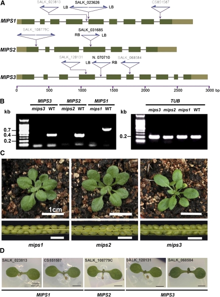
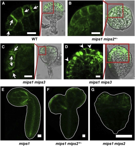
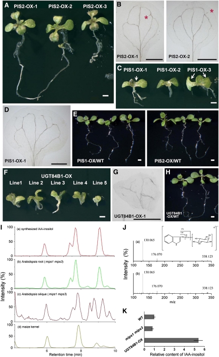
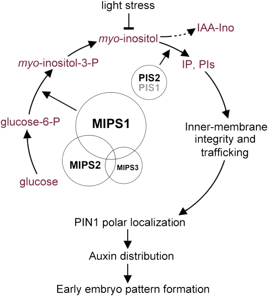
In animal cells, myo-inositol is an important regulatory molecule in several physiological and biochemical processes, including signal transduction and membrane biogenesis. However, the fundamental biological functions of myo-inositol are still far from clear in plants. Here, we report the genetic characterization of three Arabidopsis thaliana genes encoding D-myo-inositol-3-phosphate synthase (MIPS), which catalyzes the rate-limiting step in de novo synthesis of myo-inositol. Each of the three MIPS genes rescued the yeast ino1 mutant, which is defective in yeast MIPS gene INO1, and they had different dynamic expression patterns during Arabidopsis embryo development. Although single mips mutants showed no obvious phenotypes, the mips1 mips2 double mutant and the mips1 mips2 mips3 triple mutant were embryo lethal, whereas the mips1 mips3 and mips1 mips2⁺/⁻ double mutants had abnormal embryos. The mips phenotypes resembled those of auxin mutants. Indeed, the double and triple mips mutants displayed abnormal expression patterns of DR5:green fluorescent protein, an auxin-responsive fusion protein, and they had altered PIN1 subcellular localization. Also, membrane trafficking was affected in mips1 mips3. Interestingly, overexpression of PHOSPHATIDYLINOSITOL SYNTHASE2, which converts myo-inositol to membrane phosphatidylinositol (PtdIns), largely rescued the cotyledon and endomembrane defects in mips1 mips3. We conclude that myo-inositol serves as the main substrate for synthesizing PtdIns and phosphatidylinositides, which are essential for endomembrane structure and trafficking and thus for auxin-regulated embryogenesis.


Figures

Figure 1.
Figure 1.
Complementation of the Yeast ino1 Mutant. Expression of each of the three Arabidopsis MIPS complements the inositol auxotrophy phenotype of the yeast ino1 mutant. The yeast cells transformed with INO1 or empty vector were used as positive and negative controls, respectively. Plates were spotted with 10-fold serial dilutions and incubated at 30°C for 2 d.
Figure 2.
Figure 2.
Relative Expression Level of MIPS Genes in Different Arabidopsis Tissues. MIPS1, MIPS1, and MIPS3 mRNA transcript levels in 8-DAG seedlings grown on half-strength Murashige and Skoog medium and in different organs of soil-grown Arabidopsis plants were quantified by real-time PCR. The expression levels for all genes were normalized to that of TUB2. Error bars were calculated as se for three independent experiments.
Figure 3.
Figure 3.
Dynamic Expression of MIPS Genes in Seed Development. (A) to (C) GUS staining pattern of siliques from MIPS1:GUS (A), MIPS2:GUS (B), and MIPS3:GUS (C) transgenic lines. These seeds contain early torpedo stage embryos. Bars = 1 mm. (D) to (G) GUS staining of seeds from MIPS1:GUS transgenic lines in sequential developmental stages as shown at the bottom of the figure. Bars = 100 μm. (H) to (K) GUS staining of seeds from pMIPS2:GUS transgenic lines in sequential developmental stages. Bars = 100 μm. (L) to (O) GUS staining of seeds from pMIPS3:GUS transgenic lines in sequential developmental stages. Bars = 100 μm.
Figure 4.
Figure 4.
Expression and Phenotype Analysis of T-DNA Insertion Mutants for Each MIPS Gene. (A) Gene structures of MIPS1, MIPS2, and MIPS3 with the location of T-DNA insertions. The left border of each insertion was confirmed by sequencing. Exons (dark-green boxes indicate coding regions; lighter boxes indicate 5′ [left] and 3′ [right] untranslated regions) and T-DNA insertion sites (purple arrows) are indicated. SALK lines written in black are those used in further analysis and crossing. SALK lines written in gray are used in phenotype confirmation. LB, T-DNA left border; RB, T-DNA right border. Gene organizations are depicted in proportion. (B) Transcript levels of MIPS1, MIPS2, MIPS3 (left panel), and the internal control TUB2 (right panel) in wild-type (WT) and three mips T-DNA insertion mutants were determined using RT-PCR (32 cycles). (C) The 20-DAG seedling (top row) and seed (bottom row) phenotypes of mips1 (left panels), mips2 (middle panels), and mips3 (right panels) single knockout mutants under the 16-h-light and 8-h-dark optimal growth condition. Bars = 1 cm. (D) Normal cotyledon phenotype in different homozygous mips single mutants. T-DNA insertion lines for three MIPS genes were grown under the long-day light (120 μmol ·m−2 ·s−1) optimal condition. The T-DNA insertion sites and the genotypes were confirmed by PCR. Bars = 1mm.
Figure 5.
Figure 5.
The Phenotypes of mips1 under Different Light Intensity Conditions. (A) Rosette leaf phenotypes of mips1 (SALK_023626) grown under different light intensity conditions in comparison with the wild type (WT). Yellow circles indicate lesion-mimic patches. Bars = 0.5 cm. (B) Cotyledon phenotypes of the seeds from mips1 (SALK_023626) grown under different light intensity conditions. Each pie chart shows the percentage of the cotyledon phenotype proportion. Lime sectors represent the normal cotyledons and yellow sectors represent the abnormal cotyledons. Bars = 1 mm.
Figure 6.
Figure 6.
Phenotypic Analysis of mips Double Mutant Cotyledons. (A) to (H) Cotyledon phenotypes of wild-type (WT; [A]), mips1 mips2+/− ([B] to [D]), mips1 mips3 ([E] to [G]), and the complementation line of mips1 mips3 with genomic MIPS1 gene driven by its own promoter (H). Bars = 1 mm. (I) to (K) The vascular patterns of cotyledons of mips1 mips2+/− (I) and mips1 mips3 ([J] and [K]). Red asterisks in (I) indicate the paralleled veins in cotyledon petioles, and the asterisk in (J) indicates irregular vein cluster. The arrow in (I) indicates the open end of a vascular bundle and in (J) indicates the isolated vascular segment. Cotyledon fusion site of mips1 mips3 seedlings is indicated by arrow in (K). P, petiole. Bars = 500 μm. (L) to (N) Pavement cell and stomatal guard cell morphology of the wild-type (L), mips1 mips2+/− (M), and mips1 mips3 (N) double mutant cotyledons. Extremely large guard cells (arrows) and small guard cells (red arrowheads) were found in mips double mutant cotyledons. Bars = 50 μm. (O) Relative cell ploidy ratio of the wild-type and mips1 mips3 cotyledons. More than 10,000 nuclei were counted for each sample. (P) myo-inositol content in mature seeds of wild-type and mips double mutants. Asterisks indicate P value <0.001. Error bars were calculated as se for three independent experiments.
Figure 7.
Figure 7.
Embryo Development of the Wild Type and mips Double and Triple Mutants. (A) to (L) Nomarski images of cleared Arabidopsis seeds from siliques of the wild type (WT; [A] to [D]), mips1 mips2+/− ([E] to [H]), and mips1 mips3 ([I] to [L]) at the four developmental stages noted at the top of the figure. Arrows in (E) indicate the abnormal cell division orientation, and red arrowheads in (F) indicate three cotyledon primordia. The arrow pair in (K) indicates the abnormal bulges in hypocotyl. C, cotyledon primordium; H, hypocotyl; Er, embryonic root. Bars = 20 μm. (M) to (O) Dissected siliques of the wild type (M), mips1 mips2+/− (N), and mips1+/− mips2 mips3 (O). Red asterisks indicate the flat white seeds that are distinguishable from the adjacent round green seeds. (P) to (S) Nomarski images of cleared Arabidopsis seeds from mips1+/− mips2 mips3 siliques. In one silique, although most of the embryos reached the heart stage (Q), about a quarter of the embryos remained at the globular stage (P). When most of the embryos reached the torpedo stage (S), about a quarter of the embryos from the same silique remained at the globular stage (R). Bars = 20 μm.
Figure 8.
Figure 8.
Auxin Maxima Distribution in the Wild-Type, mips Double, and Triple Mutant Embryos. (A) to (C) DR5:GFP distribution in the wild-type (WT) embryos at the globular stage (A), the late heart stage (B), and the mature stage (C). (D) to (F) DR5:GFP distribution in embryos dissected from mips1 mips2+/− siliques at the globular stage (D), the late heart stage (E), and the mature stage (F). Arrow in (D) indicates the extra GFP signal in suspensor cells. (G) to (I) DR5:GFP distribution in mips1 mips3 embryos at the globular stage (G), the late heart stage (H), and the torpedo stage (I). The dashed line frame in (G) indicates the hypophysis cells with decreased DR5:GFP signals. (J) to (L) DR5:GFP distribution in embryos dissected from the flat white seeds of mips1+/− mips2 mips3 mature silique ([J] and [K]). Red arrowhead in (L) indicates the abnormal bulge in shoot apical meristem. Bars = 25 μm in (A) to (L).
Figure 9.
Figure 9.
PIN1 Localization in the Wild-Type and mips Double Mutant Embryos. (A) PIN1:GFP signals in a globular stage embryo of the wild type (WT). Arrows show the PIN1:GFP polar localization, which indicates the direction of auxin flux toward the differentiating cotyledon primordium. (B) A globular shape embryo dissected from a silique of mips1 mips2+/− showing evenly distributed PIN1:GFP signals on plasma membrane. (C) and (D) PIN1:GFP signals in globular-stage embryo of mips1 mips3. Arrows in (C) indicate the disorganized PIN1 localization on lateral plasma membranes. Arrowheads in (D) indicate PIN proteins facing outwards. In (A) to (D), the left panels show the fluorescence image of the area in the red rectangle in the right Nomarski image. Bars = 5 μm in (A) to (D). (E) to (G) Three phenotypically distinct embryos dissected from the same silique of mips1 mips2+/− showing different PIN1:GFP signals. The predicted genotypes are amips1 (E), mips1 mips2+/− (F), and mips1 mips2 (G). Bars = 25 μm.
Figure 10.
Figure 10.
Phenotypic Analysis and Biochemical Characterization of PIS2-, PIS1-, and UGT84B1-Overexpressor Lines in the mips1 mips3 Background. (A) to (H) The 12-DAG PIS2-OX (A), PIS1-OX (C), and UGT84B1-OX (F) transgenic lines in the mips1 mips3 background. The T1 generation lines shown above were selected by growing on half-strength Murashige and Skoog agar plate containing 50 μg/mL hygromycin. PIS2-OX lines in (A) show regularly symmetry two cotyledons. Arrow (left) in (C) indicates the broader cotyledon petiole; arrow pair (right) in (C) indicates the two asymmetric cotyledons. Cotyledon venations of PIS2-OX (B), PIS1-OX (D), and UGT84B1-OX (G) transgenic lines. Red asterisks in (B) indicate the extra vascular structures. Wild-type Arabidopsis overexpressing PIS2 (PIS2-OX) or PIS1 (PIS1-OX) (E) or UGT84B1 (UGT84B1-OX) (H) are also shown. Bars = 1 mm. (I) Liquid chromatography–MS chromatograms of IAA-myo-inositol quantification assay using synthesized IAA-myo-inositol (a), mips1 mips3 root (b), mips1 mips3 silique (c), and maize kernel (d). Four peaks in chromatogram (a) show four isomers of IAA-myo-inositol. (J) MS2 spectra of the first isomer (retention time, 5.9 min) in synthesized IAA-myo-inositol (a) and wild-type Arabidopsis root (b). The fraction of m/z 130 and m/z 176, as well as the [M+H]+ m/z 338.123, confirmed the IAA-myo-inositol structure of the first isomer. (K) Relative content of IAA-myo-inositol in Arabidopsis root tissue of different genotypes. Duplicate experiments were performed for each genotype. Error bars show se. WT, wild type.
Figure 11.
Figure 11.
Endomembrane of the Wild-Type and mips1 mips3 Embryo Cells. (A) to (F) Arabidopsis wild-type (WT; [A] to [C]) and mips1 mips3 double mutant ([D] to [F]) embryo cells treated with the endocytosis marker AM1-43. More than 20 siliques were analyzed for each treatment. Bars = 10 μm. (G) Endomembrane system of a wild-type embryo cell at the torpedo stage. Arrowhead indicates a double membrane bound vesicle; arrow indicates a budding vesicle. ER, endoplasmic reticulum. Bar = 200 nm. (I) Endomembrane system of an mips1 mips3 embryo cell at the torpedo stage. MT, mitochondria. Bar = 200 nm. (K) Endomembrane system of a torpedo embryo cell from PIS2-OX–transformed mips1 mips3 plant; arrow indicates a trafficking vesicle. Bar = 200 nm. (H), (J), and (L) Magnification of rectangle regions in (G), (I), and (K), respectively. Arrows in (H) and (L) indicate the single membrane bound trafficking vesicles, which were not found in (J).
Figure 12.
Figure 12.
A Proposed Model for the Biological Roles of the Three MIPS Genes in Arabidopsis Embryo Pattern Formation. The myo-inositol produced by sequential reactions, in which MIPS enzymes catalyze the rate-limiting step, can be either conjugated to IAA or converted to membrane component PtdIns. Sufficient PtdIns is important for endomembrane structure and trafficking and, thus, for auxin transport and localization to regulate proper embryo development.

References

    1. Abreu E.F.M., Aragão F.J.L. (2007). Isolation and characterization of a myo-inositol-1-phosphate synthase gene from yellow passion fruit (Passiflora edulis f. flavicarpa) expressed during seed development and environmental stress. Ann. Bot. (Lond.) 99: 285–292 - PMC - PubMed
    1. Aida M., Ishida T., Fukaki H., Fujisawa H., Tasaka M. (1997). Genes involved in organ separation in Arabidopsis: An analysis of the cup-shaped cotyledon mutant. Plant Cell 9: 841–857 - PMC - PubMed
    1. Alberti S., Gitler A.D., Lindquist S. (2007). A suite of Gateway cloning vectors for high-throughput genetic analysis in Saccharomyces cerevisiae. Yeast 24: 913–919 - PMC - PubMed
    1. Altschul S.F., Gish W., Miller W., Myers E.W., Lipman D.J. (1990). Basic local alignment search tool. J. Mol. Biol. 215: 403–410 - PubMed
    1. Bachhawat N., Mande S.C. (2000). Complex evolution of the inositol-1-phosphate synthase gene among archaea and eubacteria. Trends Genet. 16: 111–113 - PubMed

Publication types

MeSH terms