. 2011 Jun 15;71(12):4117-29.
doi: 10.1158/0008-5472.CAN-11-0176. Epub 2011 Apr 19.

Common and overlapping oncogenic pathways contribute to the evolution of acute myeloid leukemias

Affiliations

Common and overlapping oncogenic pathways contribute to the evolution of acute myeloid leukemias

Brynn T Kvinlaug et al. Cancer Res. .

Abstract

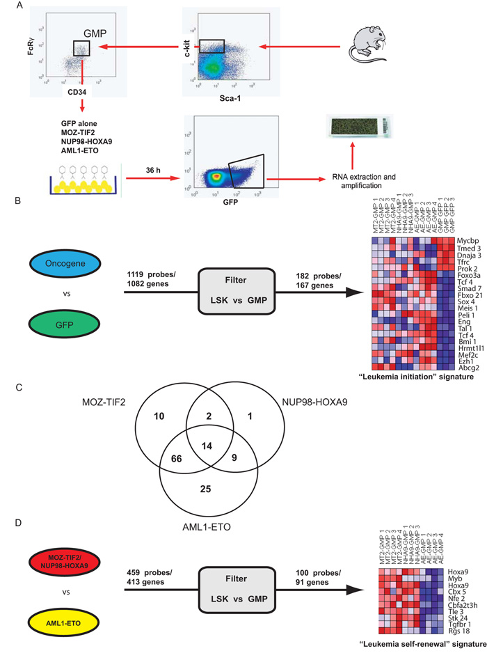
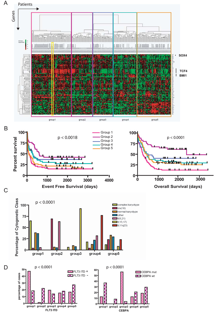
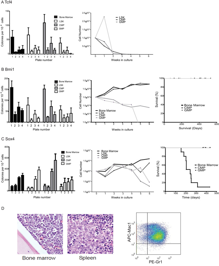
Fusion oncogenes in acute myeloid leukemia (AML) promote self-renewal from committed progenitors, thereby linking transformation and self-renewal pathways. Like most cancers, AML is a genetically and biologically heterogeneous disease, but it is unclear whether transformation results from common or overlapping genetic programs acting downstream of multiple mutations or by the engagement of unique genetic programs acting cooperatively downstream of individual mutations. This distinction is important, because the involvement of common programs would imply the existence of common molecular targets to treat AML, no matter which oncogenes are involved. Here we show that the ability to promote self-renewal is a generalized property of leukemia-associated oncogenes. Disparate oncogenes initiated overlapping transformation and self-renewal gene expression programs, the common elements of which were defined in established leukemic stem cells from an animal model as well as from a large cohort of patients with differing AML subtypes, where they strongly predicted pathobiological character. Notably, individual genes commonly activated in these programs could partially phenocopy the self-renewal function of leukemia-associated oncogenes in committed murine progenitors. Furthermore, they could generate AML following expression in murine bone marrow. In summary, our findings reveal the operation of common programs of self-renewal and transformation downstream of leukemia-associated oncogenes, suggesting that mechanistically common therapeutic approaches to AML are likely to be possible, regardless of the identity of the driver oncogene involved.


Figures

Figure 1
Figure 1. Transcription factor fusions associated with AML commonly alter self-renewal properties
A) Committed progenitors (CMP, GMP and MEP) transduced with FLT3-ITD do not serially replate in cytokine supplemented methycellulose (left panel). The right panel shows a Kaplan-Meier graph for survival of transplant recipients of FLT3-ITD transduced CMP, GMP and MEP. All animals were healthy until the time of sacrifice (up to 350 days) with no evidence of leukemia or reconstitution by the transplanted cells. B) CMP and GMP transduced with AML1-ETO serially replate in cytokine supplemented methylcellulose and grow in liquid culture (left and middle panels). Following transplantation of these populations no reconstitution was detected (right panel). C) Similar analysis is presented for NUP98-HOXA9, demonstrating that CMP, GMP and MEP transduced with NUP98-HOXA9 serially replate in cytokine supplemented methylcellulose and grow in liquid culture (upper left and middle panels). However, and in contrast to AML1-ETO, NUP98-HOXA9 transduced progenitor populations generate acute leukemias as demonstrated in the Kaplan-Meier graph in the right upper panel and in the lower panels, where representative photomicrographs of peripheral blood, bone marrow, liver and spleen demonstrate significant infiltration with leukemic blasts on a background of myeloproliferation.
Figure 2
Figure 2. Identification of immediate leukemia-associated signatures downstream of multiple AML fusion genes
A) Panel A demonstrates a schema of the experimental strategy used to identify the leukemia-associated signatures. B) Identification and prioritization of genes to generate the 167 gene (182 probe) immediate “leukemia initiation” signature. Initially the expression profiles for multiple replicates of MOZ-TIF2, AML1-ETO and NUP98-HOXA9 transduced GMPs were compared in a single two-class comparison to GFP transduced GMPs. 1119 genes were commonly dysregulated to an FDR significance level of 0.05. A filter comparing expression of the 1119 genes between normal HSC and GMP was then applied, with genes which were significantly and coordinately differentially expressed (p < 0.05) retained. This prioritized 167 genes to generate our signature. Expression patterns of 20 representative genes are presented in the left part of the panel (all genes are listed in Table S1). C) Three single two-class comparisons were also made between each of MOZ-TIF2, AML1-ETO and NUP98-HOXA9 transduced GMPs and GFP transduced GMPs and genes differentially expressed (p<0.05) were overlapped with our leukemia initiation signature. The Venn-diagram details the number of genes dysregulated by each oncogene and demonstrates significant common and overlapping pathways downstream of the three oncogenes. D) Identification and prioritization of genes to generate the 91 gene (100 probe) immediate “leukemia self-renewal” signature. The expression profiles for multiple replicates of MOZ-TIF2 and NUP98-HOXA9 transduced GMPs were compared in a single two-class comparison to AML1-ETO transduced GMPs. 413 genes were commonly dysregulated (p < 0.01). This geneset was then similarly filtered to generate our immediate leukemia self-renewal signature. Expression patterns of 10 representative probes (9 genes) are presented in the left part of the panel (all genes are listed in Table S2).
Figure 3
Figure 3. MOZ-TIF2-associated murine leukemias recapitulate an LSC hierarchy and demonstrate the evolutionary nature of LSC transcriptional programs
A) The experimental schema used to establish an LSC hierarchy for MOZ-TIF2-associated murine leukemias. Briefly, the efficiency with which leukemia was transferred to sublethally irradiated syngeneic recipients was assessed at limiting dilution as is shown for three separate populations. These were defined according to normal myeloid ontogeny and comprised a “mature” population (left panels), a “bulk” leukemia population (middle panels) and an “immature” population with the same surface phenotype as GMP (right panels). The efficiency of transfer and leukemia stem cell frequency as calculated by Poisson statistics for each population is shown below the Kaplan-Meier graphs in the lower row of panels. Greater than 3 log enrichment for leukemia stem cell frequency was demonstrated between the immature “leukemic” GMP (L-GMP) and “mature” (GFP+, Mac+ and Gr1+) populations. B) Gene expression profiles from the L-GMP population, significantly enriched for LSC activity, were then compared with their normal GMP counterpart. Expression data for a representative 20 Probes (19 genes) are shown. This demonstrates that certain genes, such as Bmi1, Meis1, Sox4, Tcf4, Hoxa9 and Smad7 remain significantly upregulated between our immediate signatures and established leukemia stem cells. However, in addition, a number of new genes widely implicated in leukemogenesis become upregulated during leukemic evolution, including Hoxa10, Hoxa7, Runx1, Lmo2 and Ctnna1.
Figure 4
Figure 4. The immediate leukemia initiation signature is present across many human AML samples and predicts for survival and disease biology
A) Sixty-seven/84 genes (80%) from the immediate leukemia initiation signature were differentially expressed across a cohort of 253 AML patients as shown in the heatmap. The geneset was able to classify this cohort into five groups using unsupervised clustering. The immediate leukemia initiation signature (outlined in the yellow box) is representative of, and tightly segregates with, group 1 when included in this analysis. B) Significant differences in survival for the five groups are demonstrated in this Kaplan-Meier estimation of event-free (p < 0.0018, left panel) and overall survival (p < 0.0001, right panel), with group 1 associated with the poorest prognosis. C) Significant differences in cytogenetic characteristics were demonstrated for individuals in each of the groups (p < 0.0001), with group 1 patients associated with a complex karyotype (poor prognosis) and a lack of rearrangements of core-binding factors and the retinoic acid receptor alpha (RARA) (good prognosis). D) Significant differences in molecular prognostic factors such as mutational status for FLT3-ITD (left panel) and CEBPA (right panel) were also noted (each p < 0.0001). Patients in group 1 demonstrated an increased incidence of the poor prognosis FLT3-ITD mutations and a lower incidence of good prognosis CEBPA mutations (see also Figures S1, 2 and 3 and supplementary Table 3).
Figure 5
Figure 5. Individual genes in the leukemia inititation signature partially phenocopy AML-associated fusion genes
A) Tcf4 did not alter the properties of bone marrow, LSK or progenitors in methylcellulose (left panel) and Tcf4 cells failed to grow in liquid culture (right panel). B) The left panel demonstrates that whole bone marrow (BM), LSK and myeloid progenitors (CMP and GMP) transduced with Bmi1 serially replate in methylcellose cultures. Both whole BM and LSK transduced with Bmi1 continue to grow in cytokine supplemented liquid culture, while growth of CMP and GMP populations is limited to 4 weeks in culture before their involution (middle panel). The right panel demonstrates that whole bone marrow or progenitors transduced with Bmi1 did not generate AML following transplantation. C) The left and middle panels demonstrate similar findings for stem and progenitor populations transduced with Sox4. In addition, BM transduced with Sox4 generated leukemia with a median latency of 28 weeks (right panel). D) Representative photomicrographs of bone marrow and spleen (left and middle panels respectively) from animals with Sox4-associated AML demonstrate heavy infiltration with immature blasts. Representative flow of the GFP positive cells from these leukemias (right panel) demonstrates a Gr1intermediate/ Mac1intermediate myeloid phenotype.

References

    1. Estey E, Dohner H. Acute myeloid leukaemia. Lancet. 2006;368:1894–1907. - PubMed
    1. Ley TJ, Mardis ER, Ding L, et al. DNA sequencing of a cytogenetically normal acute myeloid leukaemia genome. Nature. 2008;456:66–72. - PMC - PubMed
    1. Bullinger L, Dohner K, Bair E, et al. Use of gene-expression profiling to identify prognostic subclasses in adult acute myeloid leukemia. N Engl J Med. 2004;350:1605–1616. - PubMed
    1. Valk PJ, Verhaak RG, Beijen MA, et al. Prognostically useful gene-expression profiles in acute myeloid leukemia. N Engl J Med. 2004;350:1617–1628. - PubMed
    1. Jemal A, Siegel R, Ward E, et al. Cancer statistics, 2008. CA Cancer J Clin. 2008;58:71–96. - PubMed

Publication types

MeSH terms

Substances