Expression Pattern of the Pro-apoptotic Gene PAR-4 During the Morphogenesis of MCF-10A Human Mammary Epithelial Cells
- PMID: 21505560
- PMCID: PMC3047628
- DOI: 10.1007/s12307-010-0059-y
Expression Pattern of the Pro-apoptotic Gene PAR-4 During the Morphogenesis of MCF-10A Human Mammary Epithelial Cells
Abstract
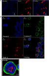
The histological organization of the mammary gland involves a spatial interaction of epithelial and myoepithelial cells with the specialized basement membrane (BM), composed of extra-cellular matrix (ECM) proteins, which is disrupted during the tumorigenic process. The interactions between mammary epithelial cells and ECM components play a major role in mammary gland branching morphogenesis. Critical signals for mammary epithelial cell proliferation, differentiation, and survival are provided by the ECM proteins. Three-dimensional (3D) cell culture was developed to establish a system that simulates several features of the breast epithelium in vivo; 3D cell culture of the spontaneously immortalized cell line, MCF10A, is a well-established model system to study breast epithelial cell biology and morphogenesis. Mammary epithelial cells grown in 3D form spheroids, acquire apicobasal polarization, and form lumens that resemble acini structures, processes that involve cell death. Using this system, we evaluated the expression of the pro-apoptotic gene PAWR (PKC apoptosis WT1 regulator; also named PAR-4, prostate apoptosis response-4) by immunofluorescence and quantitative real time PCR (qPCR). A time-dependent increase in PAR-4 mRNA expression was found during the process of MCF10A acinar morphogenesis. Confocal microscopy analysis also showed that PAR-4 protein was highly expressed in the MCF10A cells inside the acini structure. During the morphogenesis of MCF10A cells in 3D cell culture, the cells within the lumen showed caspase-3 activation, indicating apoptotic activity. PAR-4 was only partially co-expressed with activated caspase-3 on these cells. Our results provide evidence, for the first time, that PAR-4 is differentially expressed during the process of MCF10A acinar morphogenesis.
Keywords: Apoptosis; Breast cancer; Gene expression; MCF10A; PAR-4; Three dimensional (3D) cell culture.
Figures


Similar articles
-
Building bridges toward invasion: tumor promoter treatment induces a novel protein kinase C-dependent phenotype in MCF10A mammary cell acini.PLoS One. 2014 Mar 5;9(3):e90722. doi: 10.1371/journal.pone.0090722. eCollection 2014. PLoS One. 2014. PMID: 24599099 Free PMC article.
-
Tightly controlled MRTF-A activity regulates epithelial differentiation during formation of mammary acini.Breast Cancer Res. 2017 Jun 7;19(1):68. doi: 10.1186/s13058-017-0860-3. Breast Cancer Res. 2017. PMID: 28592291 Free PMC article.
-
Down-regulation of the candidate tumor suppressor gene PAR-4 is associated with poor prognosis in breast cancer.Int J Oncol. 2010 Jul;37(1):41-9. doi: 10.3892/ijo_00000651. Int J Oncol. 2010. PMID: 20514395
-
Lumen formation during mammary epithelial morphogenesis: insights from in vitro and in vivo models.Cell Cycle. 2008 Jan 1;7(1):57-62. doi: 10.4161/cc.7.1.5150. Epub 2007 Oct 9. Cell Cycle. 2008. PMID: 18196964 Review.
-
Application of the D492 Cell Lines to Explore Breast Morphogenesis, EMT and Cancer Progression in 3D Culture.J Mammary Gland Biol Neoplasia. 2019 Jun;24(2):139-147. doi: 10.1007/s10911-018-09424-w. Epub 2019 Jan 25. J Mammary Gland Biol Neoplasia. 2019. PMID: 30684066 Review.
Cited by
-
Met receptor acts uniquely for survival and morphogenesis of EGFR-dependent normal mammary epithelial and cancer cells.PLoS One. 2012;7(9):e44982. doi: 10.1371/journal.pone.0044982. Epub 2012 Sep 13. PLoS One. 2012. PMID: 23028720 Free PMC article.
-
HOXB7 Overexpression Leads Triple-Negative Breast Cancer Cells to a Less Aggressive Phenotype.Biomedicines. 2021 May 5;9(5):515. doi: 10.3390/biomedicines9050515. Biomedicines. 2021. PMID: 34063128 Free PMC article.
References
-
- Petersen OW, Rønnov-Jessen L, Howlett AR, Bissell MJ. Interaction with basement membrane serves to rapidly distinguish growth and differentiation pattern of normal and malignant human breast epithelial cells. Proc Natl Acad Sci USA. 1992;89(19):9064–9068. doi: 10.1073/pnas.89.19.9064. - DOI - PMC - PubMed
LinkOut - more resources
Full Text Sources
Research Materials
