. 2011 Jan-Mar;4(1):63-74.
Epub 2011 Feb 25.

The role of confocal microscopy in the dermato-oncology practice

Affiliations

The role of confocal microscopy in the dermato-oncology practice

A Diaconeasa et al. J Med Life. 2011 Jan-Mar.

Abstract

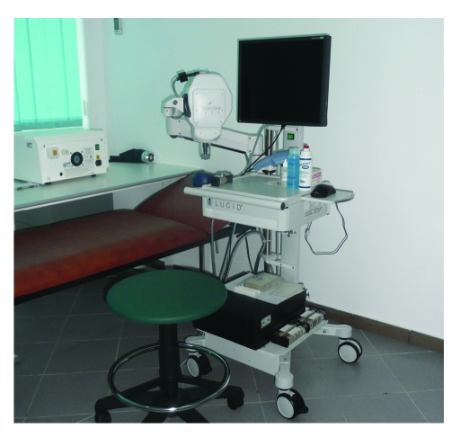
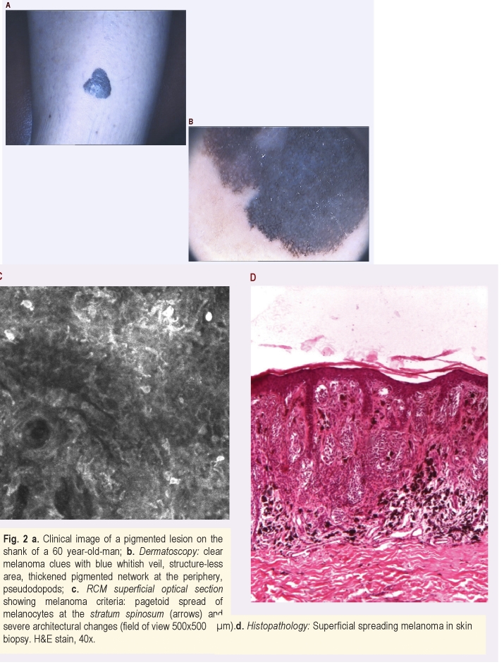
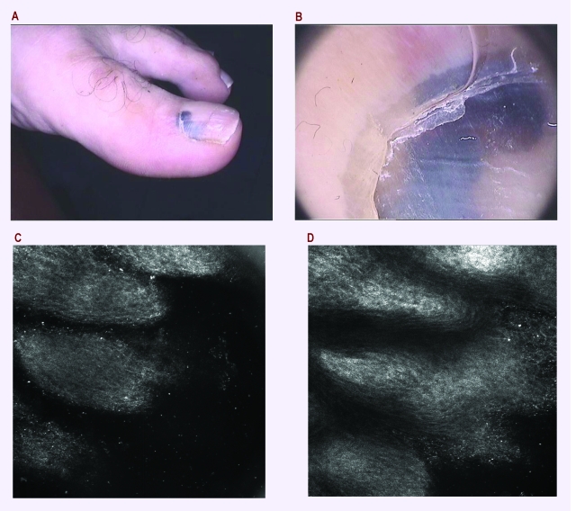
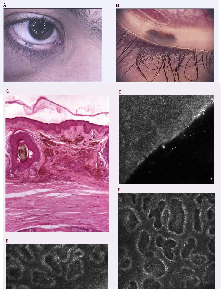
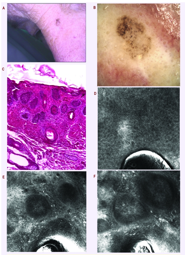
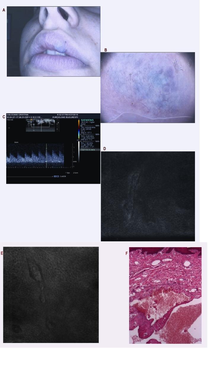
Reflectance-mode confocal microscopy (RCM) is a new in vivo skin imaging technique. We present our one-year experience in RCM examinations in skin tumors and the retrospective analysis of patients enrolled in the Dermatological Department of 'N. Paulescu' Institute using the Fotofinder Dermoscope IIŴ for the dermatoscopy analysis and VivaScope 1500Ŵ for in vivo RCM. We established the rank of RCM in the complex algorithm of skin cancer diagnose, showing that the presented experience can open new possibilities to implement this automated image analyzing system in the routine practice. Our analyzed cases clearly showed that confocal microscopy, therefore, optical biopsy, could guide the clinician towards an accurate diagnosis before surgical removal. Moreover, we emphasized that the development of this technique increases the potential of future teledermatologic applications.

Keywords: dermatoscopy; optical biopsy.


Figures

Figure 1
Figure 1
Reflectance–mode confocal microscopy VivaScope 1500
Figure 2
Figure 2
Clinical image of a pigmented lesion on the shank of a 60 year–old–man; b. Dermatoscopy: clear melanoma clues with blue whitish veil, structure–less area, thickened pigmented network at the periphery, pseudodopods; c. RCM superficial optical section showing melanoma criteria: pagetoid spread of melanocytes at the stratum spinosum (arrows) and severe architectural changes (field of view 500x500 ॖm).d. Histopathology: Superficial spreading melanoma in skin biopsy. H and E stain, 40x.
Figure 3
Figure 3
a. Dermatoscopy of a pigmented lesion in a patient with a history of melanoma: few irregular dots and globules corresponding to melanocytic nests seen on pathology, no clues for melanoma though; b. Histopathology: Junctional dysplastic nevus with architectural and cytological atypia; there is a mild superficial perivascular lymphocytic infiltration. H and E stain, 100x. c. RCM superficial optical section showing regularly arranged uniform keratinocytes in a honeycomb pattern in stratum granulosum (500x500 ॖm). d. RCM deeper optical section with edged papillae at dermal–epidermal junction; RCM images suggest a benign lesion.
Figure 4
Figure 4
a. Clinical aspect of a recently appeared pigmented lesion on the calf of the leg of a 15–year–old girl; b. Dermatoscopy: structureless area, few peripheric structures; possible Reed nevus with heavily pigmented cells, Clark nevus with compactly aggregated nest of melanocytes; blue nevus or melanoma cannot be excluded; c. Histopathology: Lentiginous junctional nevus; single cells and nests of nevus cells are arranged at the tips of rete ridges; the upper dermis contains an infiltration of melanophages and mononuclear cells. H and E stain, 40x. d. RCM superficial optical section showing few pagetoid cells (arrows); e. RCM deeper optical section with normal stratum basale; f. RCM deep optical section with edged papillae (arrows) and normal melanocytes nests, suggesting a nevus.
Figure 5
Figure 5
a. Clinical aspect of a nail lesion in a 55–year–old man; b. Dermatoscopy: pseudo–Hutchinson sign, no clear clues for melanoma–possible hemorrhage; c. RCM superficial optical section of the nail fold with normal stratum corneum and no atypical cells (500x500 microm); d. Deeper RCM image showing no sign of melanoma, suggesting a subungual hemorrhage (as confirmed after surgery).
Figure 6
Figure 6
a. Pigmented lesion of the eyelid in a 17–year–old boy; b. Dermatoscopy: unspecific dermatoscopic pattern due to the particular location; if we are following the classical pattern analysis we can differentiate 2 colors, irregularly distributed, few dots in the periphery and also some peripheral linear structures, so it is impossible to exclude a melanoma after this assessment; c. Histopathology: Intradermal nevus; the upper dermis contains nests of nevus cells and a moderate amount of melanin. H and E stain, 40x. d. RCM superficial optical section showing a normal stratum spinosum (500x500 µm); e. Deeper RCM image with edged papillae (as a sign of a benign lesion); f. Typical RCM aspect of a nevus with melanin providing strong cytoplasmic contrast at dermal–epidermal junction.
Figure 7
Figure 7
a. Pigmented lesion on the face of a women aged 65; b. Dermatoscopy: very suspicious lesion for melanoma with annular granular pattern, dots invading the follicular openings, pseudo pigmented thickened network; no clue for BCC despite the clinical aspect, no arborising vessels, nor blue gray ovoid nests; possible lentigo maligna melanoma, pigmented actinic keratosis or lichen planus–like actinic keratosis; c. Histopathology: Nodular basal cell carcinoma; nestsof tumor cells show peripheral palisading. H and E stain, 40x. d. RCM superficial optical section showing a normal stratum spinosum (500x500 micro m); e. Deeper RCM optical section revealing nodular type basal cell carcinoma with palisading and clefting (arrow); f. RCM image showing inflammatory infiltrates and leukocyte adhesion and rolling (arrows); RCM suggests a diagnosis of basal cell carcinoma.
Figure 8
Figure 8
a. Clinical aspect of a blue tumor on the upper lip in a women aged 21; b. Dermatoscopy: red and blue lagoons–specific pattern for a vascular structure; c. Ultrasonography examination suggesting a solid tumor; after this examination the surgical excision is decided; d and e. RCM images showing capillary loops and still suggesting a vascular lesion (500x500 micro micro m field of view); f. Histopathology: Dilated vascular spaces filled with erythrocytes and lined by a single layer of flattened endothelial cells and a thin wall of fibrous tissue. H and E stain, 40x.

Similar articles

Cited by

References

    1. Curiel–Lewandrowski C, Williams CM, Swindells KJ, Tahan SR, Aster S, Frankenthaler RA, Gonzalez S. Use of In Vivo Confocal Microscopy in Malignant Melanoma: an Aid in Diagnosis and Assessment of Surgical and Nonsurgical Therapeutic Approaches. Arch Dermatol. 2004;140(9):1127–1132. - PubMed
    1. Gerger A, Koller S, Weger W, Richtig E, Kerl H, Samonigg H, Krippl P. Sensitivity and Specificity of Confocal Laser–Scanning Microscopy for In Vivo Diagnosis of Malignant Skin Tumors. Cancer. 2006;107(1):193–200. - PubMed
    1. Rajadhyaksha M, Grossman M, Esterowitz D. In vivo confocal scanning laser microscopy of human skin: melanin provides strong contrast. J Invest Dermatol . 1995;104:946–952. - PubMed
    1. Diepgen TL, Mahler V. The epidemiology of skin cancer. Br J Dermatol. 2002;146:1–6. - PubMed
    1. Green A. Changing patterns of non–melanoma skin cancer. Epithelial Cell Biol . 1992;(1):47–51. - PubMed

Publication types

LinkOut - more resources