. 2011 Dec;68(23):3919-31.
doi: 10.1007/s00018-011-0684-8. Epub 2011 Apr 20.

Clusterin facilitates in vivo clearance of extracellular misfolded proteins

Affiliations

Clusterin facilitates in vivo clearance of extracellular misfolded proteins

Amy R Wyatt et al. Cell Mol Life Sci. 2011 Dec.

Abstract

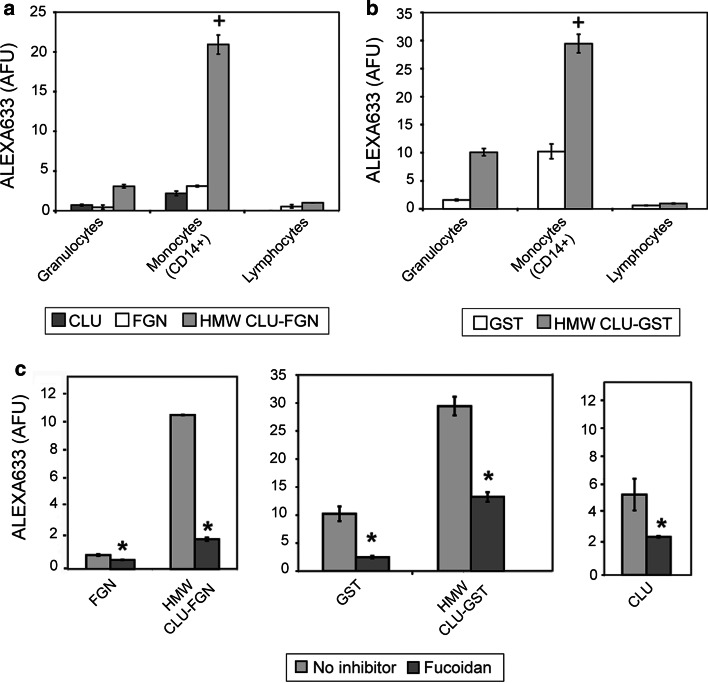
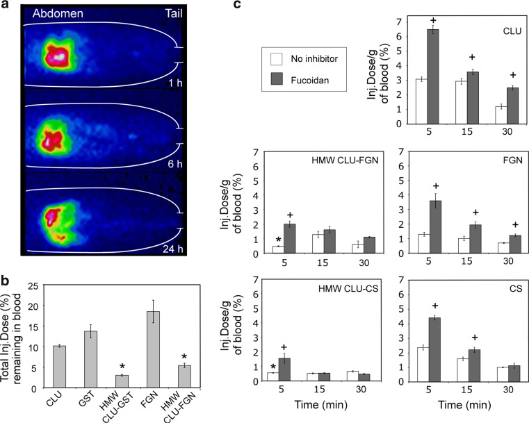
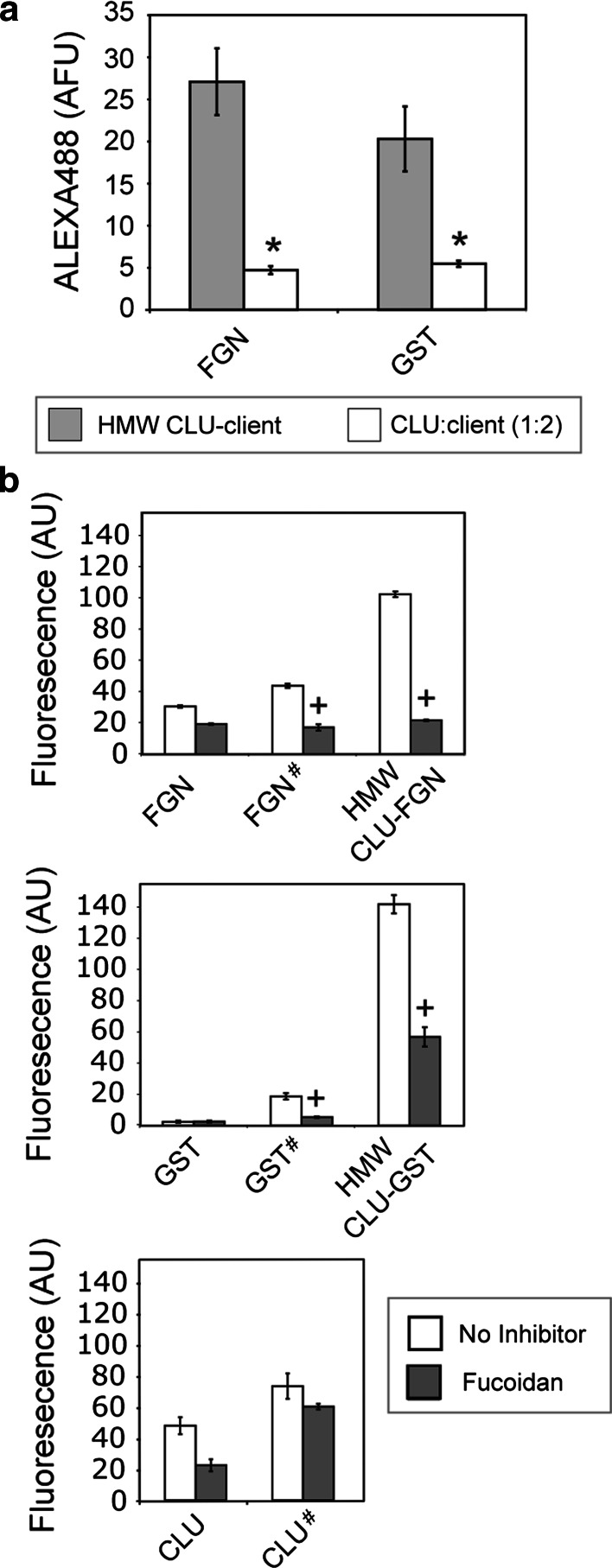
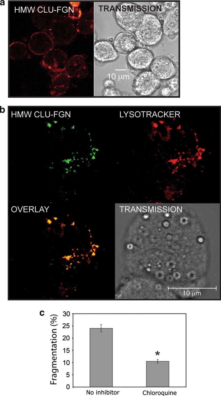
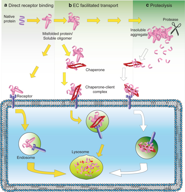
The extracellular deposition of misfolded proteins is a characteristic of many debilitating age-related disorders. However, little is known about the specific mechanisms that act to suppress this process in vivo. Clusterin (CLU) is an extracellular chaperone that forms stable and soluble complexes with misfolded client proteins. Here we explore the fate of complexes formed between CLU and misfolded proteins both in vitro and in a living organism. We show that proteins injected into rats are cleared more rapidly from circulation when complexed with CLU as a result of their more efficient localization to the liver and that this clearance is delayed by pre-injection with the scavenger receptor inhibitor fucoidan. The CLU-client complexes were found to bind preferentially, in a fucoidan-inhibitable manner, to human peripheral blood monocytes and isolated rat hepatocytes and in the latter cell type were internalized and targeted to lysosomes for degradation. The data suggest, therefore, that CLU plays a key role in an extracellular proteostasis system that recognizes, keeps soluble, and then rapidly mediates the disposal of misfolded proteins.


Figures

Fig. 1
Fig. 1
Binding of HMW CLU–client complexes and control proteins to human peripheral blood leukocytes and the effect of fucoidan pre-treatment, assessed by flow cytometry. Freshly isolated human leukocytes were incubated with 100 μg/ml biotinylated a clusterin (CLU), fibrinogen (FGN) or HMW CLU–FGN, or b GST or HMW CLU–GST, and then 5 μg/ml streptavidin (SA)-Alexa Fluor® 633. In separate experiments, the binding of the same proteins to CD14+ monocytes pre-incubated with or without 500 μg/ml fucoidan was measured as described (c). The results are the geometric mean of the Alexa Fluor® 633 fluorescence in arbitrary fluorescence units (AFU) (n = 3 ± standard deviation). +, denotes significantly higher binding of HMW CLU–client complexes compared across the cell types and also compared to the binding of uncomplexed control proteins to all cell types (as shown on the same panel; Tukey HSD, and Student’s t test, p < 0.01). Asterisks denotes significant inhibition by fucoidan (Student’s t test, p < 0.05)
Fig. 2
Fig. 2
Clearance of blood-borne 123I-labeled HMW CLU–client complexes and control proteins in Sprague–Dawley rats. a SPECT imaging of the lower body of a rat (orientation indicated by the labels Abdomen and Tail) injected with 123I-HMW CLU–FGN via the tail vein. Progressively higher levels of radioactivity are indicated by the pseudocolor gradient moving from blue to green, then yellow, red, and finally white. The times shown are p.i. and the images shown are representative of three different experiments. Similar results were obtained for native and residual soluble heated CLU and FGN (not shown). b Rats were injected with 123I-labeled HMW CLU–client complex or native control proteins. The radioactivity per gram of blood was measured 1 h p.i. and the total activity was calculated by estimating the blood volume of each rat (see “Materials and methods”). Data points represent means (n = 3 ± standard deviation). c Panels showing the levels of circulating radioactivity in blood up to 30 min after rats were injected with 123I-labeled HMW CLU–client complex or uncomplexed control protein with or without pre-injection with fucoidan. Data points represent means (n = 4 ± standard deviation).+, denotes significantly higher radioactivity in the blood of rats pre-injected with fucoidan (Student’s t test, p < 0.01). Asterisks denotes significantly lower radioactivity compared to CLU and the relevant uncomplexed control proteins (Tukey HSD, p < 0.01)
Fig. 3
Fig. 3
Biodistribution at 5 min p.i. of 123I labeled HMW CLU–client protein complexes and control proteins in Sprague-Dawley rats, with and without pre-injection of fucoidan. The identity of the corresponding HMW CLU–client complexes and control proteins are indicated on each panel. Data points represent the mean percentage of the injected dose/g of tissue (n = 4 ± standard deviation) and are corrected for any radioactivity remaining in the tail. Asterisks denotes significantly reduced radioactivity in animals pre-injected with fucoidan and +, denotes significantly increased radioactivity in animals pre-injected with fucoidan (Student’s t test, p < 0.01)
Fig. 4
Fig. 4
Flow cytometric measurements of the binding of HMW CLU–client complexes and control proteins to rat liver cells. a Hepatocytes were incubated with biotinylated HMW CLU–FGN or HMW CLU–GST, or mixtures of (1) biotinylated CLU and biotinylated FGN, or (2) biotinylated CLU and biotinylated GST (at the same final mass concentrations, and with CLU:client mass ratio = 1:2 in both cases), and then incubated with SA-Alexa Fluor® 488. Asterisks indicates significantly less binding of the uncomplexed proteins compared to that of the HMW CLU–client protein complexes (Student’s t test, p < 0.01). b In separate experiments, hepatocytes were incubated with or without fucoidan before incubation with biotinylated (1) FGN, residual soluble heated FGN (FGN #) or HMW CLU–FGN, or (2) GST, residual soluble heated GST (GST#) or HMW CLU–GST, or (3) CLU or residual soluble heated CLU (CLU#), followed by SA-Alexa Fluor® 488. +, indicates significantly less binding of the uncomplexed proteins compared to the HMW CLU–client complexes (Student’s t test, p < 0.01). In all cases, the results are the geometric mean fluorescence in arbitrary units (AU; n = 3 ± standard error)
Fig. 5
Fig. 5
Binding, internalization and degradation of HMW CLU–client complexes by rat hepatocytes. a Confocal fluorescence image and the corresponding transmission image of rat hepatocytes incubated with Alexa Fluor® 488-labeled HMW CLU–FGN for 2 h on ice. b Confocal fluorescence images of hepatocytes incubated with Alexa Fluor® 488-labeled HMW CLU–FGN and Lysotracker Red DND-99 for 2 h at 37°C. Also shown are an overlay of the two fluorescence images and the corresponding transmission image (as indicated on the Fig). The scale bars are 10 μm. c Effect of the lysosomal protease inhibitor chloroquine on the degradation of internalized CLU–GST complexes by rat hepatocytes (see “Materials and methods”). Asterisks denotes significant inhibition of fragmentation by chloroquine (Student’s t test, p < 0.01)
Fig. 6
Fig. 6
Proposed mechanism for maintenance of extracellular proteostasis. Under normal physiological conditions a scavenger receptors may directly bind misfolded proteins locally. b Circulating extracellular chaperones target and bind to misfolded proteins, maintaining their solubility and facilitating their transport to scavenger receptors. c When extracellular proteostasis is disrupted, insoluble protein aggregates can form giving rise to activated proteases (e.g. plasmin). Extracellular chaperones interact with the proteolytic fragments and facilitate their transport to scavenger receptors. In all cases, delivery to scavenger receptors results in the intracellular transport of misfolded proteins to lysosomes for degradation

Similar articles

Cited by

References

    1. Dobson CM. Protein folding and misfolding. Nature. 2003;426:884–890. doi: 10.1038/nature02261. - DOI - PubMed
    1. Ben-Zvi A, Miller EA, Morimoto RI. Collapse of proteostasis represents an early molecular event in Caenorhabditis elegans aging. Proc Natl Acad Sci USA. 2009;106:14914–14919. doi: 10.1073/pnas.0902882106. - DOI - PMC - PubMed
    1. Wyatt AR, Yerbury JJ, Poon S, Wilson MR. Therapeutic targets in extracellular protein deposition diseases. Curr Med Chem. 2009;16:2855–2866. doi: 10.2174/092986709788803187. - DOI - PubMed
    1. Yerbury JJ, Stewart EM, Wyatt AR, Wilson MR. Quality control of protein folding in extracellular space. EMBO Rep. 2005;6:1131–1136. doi: 10.1038/sj.embor.7400586. - DOI - PMC - PubMed
    1. Hartl FU, Hayer-Hartl M. Molecular chaperones in the cytosol: from nascent chain to folded protein. Science. 2002;295:1852–1857. doi: 10.1126/science.1068408. - DOI - PubMed

Publication types

MeSH terms