. 2011 Sep;89(9):915-25.
doi: 10.1007/s00109-011-0758-y. Epub 2011 Apr 20.

Lafora progressive myoclonus epilepsy: NHLRC1 mutations affect glycogen metabolism

Affiliations

Lafora progressive myoclonus epilepsy: NHLRC1 mutations affect glycogen metabolism

Philippe Couarch et al. J Mol Med (Berl). 2011 Sep.

Abstract

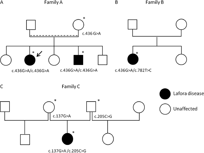
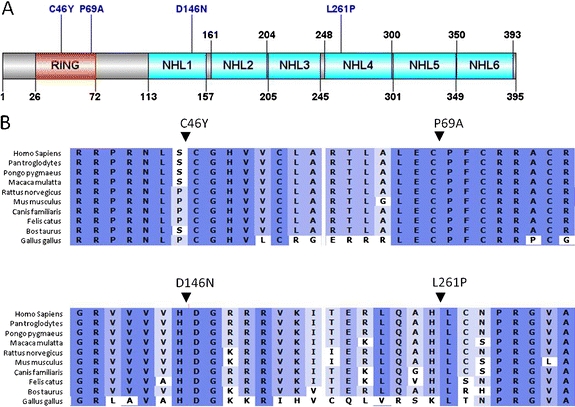
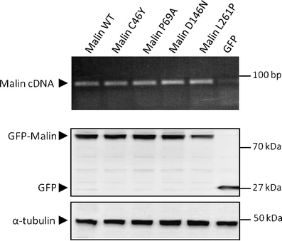
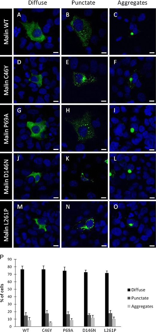
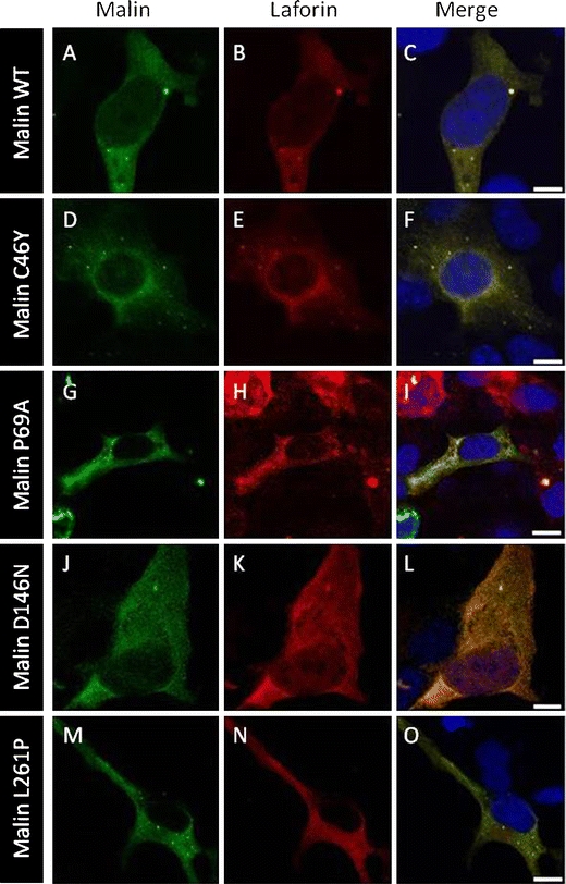
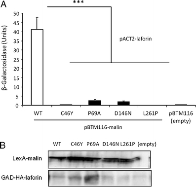
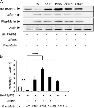
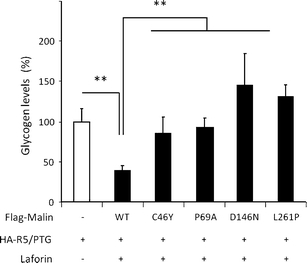
Lafora disease is a fatal autosomal recessive form of progressive myoclonus epilepsy. Patients manifest myoclonus and tonic-clonic seizures, visual hallucinations, intellectual, and progressive neurologic deterioration beginning in adolescence. The two genes known to be involved in Lafora disease are EPM2A and NHLRC1 (EPM2B). The EPM2A gene encodes laforin, a dual-specificity protein phosphatase, and the NHLRC1 gene encodes malin, an E3-ubiquitin ligase. The two proteins interact with each other and, as a complex, are thought to regulate glycogen synthesis. Here, we report three Lafora families with two novel pathogenic mutations (C46Y and L261P) and two recurrent mutations (P69A and D146N) in NHLRC1. Investigation of their functional consequences in cultured mammalian cells revealed that malin(C46Y), malin(P69A), malin(D146N), and malin(L261P) mutants failed to downregulate the level of R5/PTG, a regulatory subunit of protein phosphatase 1 involved in glycogen synthesis. Abnormal accumulation of intracellular glycogen was observed with all malin mutants, reminiscent of the polyglucosan inclusions (Lafora bodies) present in patients with Lafora disease.


Figures

Fig. 1
Fig. 1
Identification of NHLRC1 mutations. a Segregation of the c.436G>A (p.Asp146Asn/D146N) mutation in family A. Arrow indicates the index case, dashed line indicates probable consanguinity. b Detection of c.782T>C (p.Leu261Pro/L261P) and c.436G>A (p.Asp146Asn/D146N) mutations in patient of family B. c Segregation of c.137G>A (p.Cys46Tyr/C46Y) and c.205C>G (p.Pro69Ala/P69A) mutations in family C. Asterisk indicates DNA sample available for the study
Fig. 2
Fig. 2
Representation of malin showing mutations. a Schematic representation showing domain organization and location of the mutations of the malin protein. b Multiple protein alignment of malin showing conservation of Cys46, Pro69, Asp146, and Leu261 residues in different vertebrates
Fig. 3
Fig. 3
Expression levels of mutants of malin. COS-7 cells were transiently transfected with the wild-type (WT) or indicated mutant GFP-malin-expressing plasmids. Upper panel RT-PCR products of exogenous wild-type and mutants of malin separated on a 1.5% agarose gel. All mRNA transcripts were detected in comparable amounts. Bottom panel detection of wild type and mutants of malin by Western blot with anti-GFP antibody. Equal amounts of proteins were loaded as shown by the α-tubulin control
Fig. 4
Fig. 4
Subcellular distribution of malin mutants. COS-7 cells were transiently transfected with the wild-type (WT) or indicated mutant GFP-malin-expressing plasmids. Malin was detected with a confocal microscope using the GFP-derived fluorescence, and nuclei were stained with DAPI. All malin variants showed a cytoplasmic localization, either as a diffuse or punctate staining or as large aggregates. Most representative localizations of wild-type (WT) malin (ac), C46Y (df), P69A (gi), D146N (jl), and L261P (mo) mutants are shown. Magnification bars, 10 μm (p). Histograms showing the percentage of cells in each experiment (WT, n = 1,140; C46Y, n = 1,247; P69A, n = 1,262; D146N, n = 1,306; L261P, n = 1,145 cells; bars indicate SEM of three independent transfections)
Fig. 5
Fig. 5
Malin mutants colocalize with laforin. COS-7 cells were transiently cotransfected with the GFP-malin and laforin-Myc plasmids. Direct visualization of GFP-malin (green) and immunofluorescence with anti-Myc (red) was performed. Most representative localizations with wild-type (WT) malin (ac), C46Y (df), P69A (gi), D146N (jl), and L261P (mo) mutants are shown. Magnification bars, 10 μm
Fig. 6
Fig. 6
Malin mutations affect yeast two-hybrid interaction with laforin. Yeast CTY10.5d cells were cotransformed with plasmids pBTM116-malin (WT or indicated mutants) or empty vector (pBTM116) and plasmid pACT2-laforin. a Protein interaction was estimated using the yeast two-hybrid system, by measuring the β-galactosidase activity. Values correspond to means from four to six different transformants (bars indicate SEM). b Western blot analysis indicated that all proteins were expressed at similar levels
Fig. 7
Fig. 7
Malin mutations do not downregulate R5/PTG protein levels. HEK293 cells were cotransfected with pCMV-HA-R5/PTG, pCIneo-laforin, and pFLAG-Malin wild-type (WT) or different mutant plasmids. a Twenty-four hours after transfection, cell extracts (25 μg) were analyzed by Western blotting using anti-R5/PTG, anti-laforin, anti-Flag, and anti-actin antibodies. b Relative intensity of the R5/PTG bands was normalized to the actin levels and referred to the levels found in cells only expressing R5/PTG. Bars indicate SEM of three independent experiments: **p < 0.01 and ***p < 0.001
Fig. 8
Fig. 8
Malin mutations do not downregulate the glycogenic activity of R5/PTG. Glycogen levels were measured in transfected HEK293 cells with pCMV-HA-R5/PTG, pCINEO-laforin, and pFLAG-malin wild-type (WT) or different mutant plasmids. Values were referred to those found in cells expressing only HA-R5/PTG. Bars indicate SEM of three independent experiments. **p < 0.01

References

    1. Minassian BA. Lafora's disease: towards a clinical, pathologic, and molecular synthesis. Pediatr Neurol. 2001;25:21–29. doi: 10.1016/S0887-8994(00)00276-9. - DOI - PubMed
    1. Minassian BA, Lee JR, Herbrick JA, Huizenga J, Soder S, Mungall AJ, Dunham I, Gardner R, Fong CY, Carpenter S, et al. Mutations in a gene encoding a novel protein tyrosine phosphatase cause progressive myoclonus epilepsy. Nat Genet. 1998;20:171–174. doi: 10.1038/2470. - DOI - PubMed
    1. Serratosa JM, Gomez-Garre P, Gallardo ME, Anta B, de Bernabe DB, Lindhout D, Augustijn PB, Tassinari CA, Malafosse RM, Topcu M, et al. A novel protein tyrosine phosphatase gene is mutated in progressive myoclonus epilepsy of the Lafora type (EPM2) Hum Mol Genet. 1999;8:345–352. doi: 10.1093/hmg/8.2.345. - DOI - PubMed
    1. Chan EM, Young EJ, Ianzano L, Munteanu I, Zhao X, Christopoulos CC, Avanzini G, Elia M, Ackerley CA, Jovic NJ, et al. Mutations in NHLRC1 cause progressive myoclonus epilepsy. Nat Genet. 2003;35:125–127. doi: 10.1038/ng1238. - DOI - PubMed
    1. Gentry MS, Worby CA, Dixon JE. Insights into Lafora disease: malin is an E3 ubiquitin ligase that ubiquitinates and promotes the degradation of laforin. Proc Natl Acad Sci USA. 2005;102:8501–8506. doi: 10.1073/pnas.0503285102. - DOI - PMC - PubMed

MeSH terms