. 2011 Aug;63(8):2407-15.
doi: 10.1002/art.30399.

Association of endogenous anti-interferon-α autoantibodies with decreased interferon-pathway and disease activity in patients with systemic lupus erythematosus

Affiliations

Association of endogenous anti-interferon-α autoantibodies with decreased interferon-pathway and disease activity in patients with systemic lupus erythematosus

Alyssa M Morimoto et al. Arthritis Rheum. 2011 Aug.

Abstract

Objective: Numerous observations implicate interferon-α (IFNα) in the pathophysiology of systemic lupus erythematosus (SLE); however, the potential impact of endogenous anti-IFNα autoantibodies (AIAAs) on IFN-pathway and disease activity is unclear. The aim of this study was to characterize IFN-pathway activity and the serologic and clinical profiles of AIAA-positive patients with SLE.

Methods: Sera obtained from patients with SLE (n = 49), patients with rheumatoid arthritis (n = 25), and healthy control subjects (n = 25) were examined for the presence of AIAAs, using a biosensor immunoassay. Serum type I IFN bioactivity and the ability of AIAA-positive sera to neutralize IFNα activity were determined using U937 cells. Levels of IFN-regulated gene expression in peripheral blood were determined by microarray, and serum levels of BAFF, IFN-inducible chemokines, and other autoantibodies were measured using immunoassays.

Results: AIAAs were detected in 27% of the serum samples from patients with SLE, using a biosensor immunoassay. Unsupervised hierarchical clustering analysis identified 2 subgroups of patients, IFN(low) and IFN(high) , that differed in the levels of serum type I IFN bioactivity, IFN-regulated gene expression, BAFF, anti-ribosomal P, and anti-chromatin autoantibodies, and in AIAA status. The majority of AIAA-positive patients had significantly lower levels of serum type I IFN bioactivity, reduced downstream IFN-pathway activity, and lower disease activity compared with the IFN(high) patients. AIAA-positive sera were able to effectively neutralize type I IFN activity in vitro.

Conclusion: Patients with SLE commonly harbor AIAAs. AIAA-positive patients have lower levels of serum type I IFN bioactivity and evidence for reduced downstream IFN-pathway and disease activity. AIAAs may influence the clinical course in SLE by blunting the effects produced by IFNα.


Figures

Figure 1
Figure 1
Identification of endogenous anti–interferon α (IFNα) autoantibodies in sera from patients with systemic lupus erythematosus (SLE). Sera obtained from rontalizumab-naive patients with SLE (n = 32) and from healthy donors (n = 30) were tested using an enzyme-linked immunosorbent assay originally designed to detect rontalizumab, a monoclonal antibody specific for human IFNα. Signals generated by rontalizumab (0.31–2.5 μ/ml) spiked into a pool of healthy donor sera are represented by the hatched bars (controls). The double horizontal lines show the mean (+2 SD) signal generated by sera from 30 healthy donors. OD450 = optical density at 450 nm.
Figure 2
Figure 2
Detection of endogenous anti–interferon-α (IFNα) autoantibodies (AIAAs) in sera from patients with systemic lupus erythematosus (SLE), using surface plasmon resonance. Binding to human IFNα or human growth hormone by sera from patients with SLE and patients with rheumatoid arthritis (RA) was measured using a surface plasmon resonance-based immunoassay. Representative profiles of binding of AIAA-positive SLE sera to IFNα (A), AIAA-positive SLE sera to IFNα, with secondary detection using anti-human IgG antibodies (B), AIAA-negative RA sera to IFNα (C), and AIAA-positive SLE sera to human growth hormone (D) are shown. The dotted lines represent the signal generated by either IFNα (A, B, and C) or human growth hormone (D) alone (baseline); the anti-IgG baseline is the signal generated by IFNα and the anti-IgG antibody reagent. Arrows denote the times when serum was injected. Double-headed arrows represent the net amount of material bound. The arrowhead indicates the time at which anti-IgG antibodies were injected.
Figure 3
Figure 3
Reduced levels of IFN-regulated metrics, autoantibodies, and disease activity in AIAA-positive patients with SLE. A, Top, Unsupervised hierarchical clustering of analytes with decreased mean levels in AIAA-positive patients (purple) versus AIAA-negative patients (green). Two distinct clusters were revealed: IFNhigh and IFNlow. Bottom, Supervised clustering analysis of the remaining analytes and clinical parameters, showing their relative levels in patients in the IFNhigh and IFNlow groups. Data are displayed in a heat map, with grey indicating average values, blue indicating lower-than-average values, and red indicating higher-than-average values. White boxes indicate missing data. B–D, Serum type I IFN bioactivity levels, IFN signature metric (ISM) values, and serum BAFF levels (B), antiribosomal P and anti-chromatin antibody levels (C), and SLE Disease Activity Index (SLEDAI) scores (D) in the IFNhigh group (n = 21), the IFNlow/AIAA-positive group (n = 11), and the IFNlow/AIAA-negative group (n = 17). For the SLEDAI score, data were not available for 2 patients in the IFNhigh group and for 1 patient in the IFNlow/AIAA-negative group. Horizontal lines indicate the medians. Wilcoxon’s test was used to compare groups. The P values shown were adjusted for multiple testing using Benjamini and Hochberg correction. LU = luciferase units; BLC = B lymphocyte chemokine; anti-dsDNA = anti-double-stranded DNA; MCP-1 = monocyte chemoattractant protein 1; IP-10 = IFNγ-inducible 10-kd protein; I-TAC = IFN-inducible T cell α chemoattractant (see Figure 2 for other definitions).
Figure 4
Figure 4
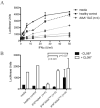
Sera from AIAA-positive patients with SLE neutralize type I IFN activity. A, U937 cells stably expressing an IFN-responsive luciferase reporter plasmid (U937-Mx1-Luc) were cultured in the presence of the indicated concentrations of recombinant IFNα and medium, healthy control sera, or sera from AIAA-positive patients with SLE. Luciferase activity was measured in cell lysates. Data are displayed as the mean ± SD for duplicate wells at each concentration. B, Peripheral blood mononuclear cells (PBMCs) from a healthy donor were incubated in the presence or absence of CL097. Following 24 hours of stimulation, supernatant was collected. U937-Mx1-Luc cells were cultured in the presence of PBMC supernatant and serum samples, as indicated. Luciferase activity was measured in cell lysates. Values for media and healthy control samples are the mean of duplicate wells. Values for the other groups are the mean ± SD. P values were determined by Mann-Whitney test. See Figure 2 for other definitions.

References

    1. Pestka S, Krause CD, Walter MR. Interferons, interferon-like cytokines, and their receptors. Immunol Rev. 2004;202:8–32. - PubMed
    1. Ronnblom L, Eloranta ML, Alm GV. The type I interferon system in systemic lupus erythematosus [review] Arthritis Rheum. 2006;54:408–20. - PubMed
    1. Hooks JJ, Jordan WG, Cuppos T, Montsopoulos TH, Fauci AS, Notkins AL. Multiple interferons in the circulation of patients with systemic lupus erythematosus and vasculitis. Arthritis Rheum. 1982;25:396–400. - PubMed
    1. Preble OT, Black RJ, Friedman RM, Klippel JH, Vilcek J. Systemic lupus erythematosus: presence in human serum of an unusual acid-labile leukocyte interferon. Science. 1982;216:429–31. - PubMed
    1. Blanco P, Palucka AK, Gill M, Pascual V, Banchereau J. Induction of dendritic cell differentiation by IFN-α in systemic lupus erythematosus. Science. 2001;294:1540–3. - PubMed

MeSH terms