. 2011 May 23;7(10):1432-9.
doi: 10.1002/smll.201002274. Epub 2011 Apr 20.

Vaults engineered for hydrophobic drug delivery

Affiliations

Vaults engineered for hydrophobic drug delivery

Daniel C Buehler et al. Small. .

Abstract

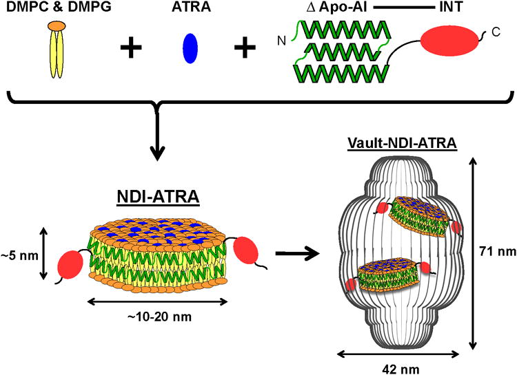
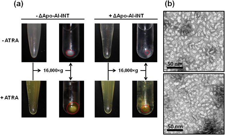
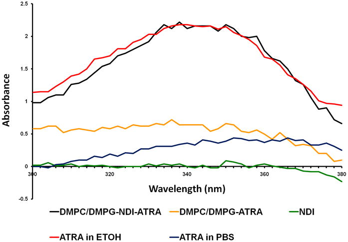
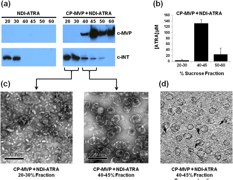
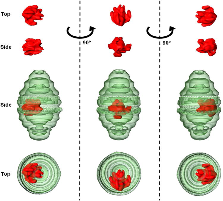
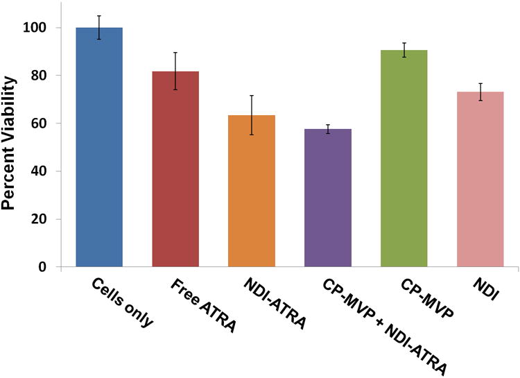
The vault nanoparticle is one of the largest known ribonucleoprotein complexes in the sub-100 nm range. Highly conserved and almost ubiquitously expressed in eukaryotes, vaults form a large nanocapsule with a barrel-shaped morphology surrounding a large hollow interior. These properties make vaults an ideal candidate for development into a drug delivery vehicle. In this study, the first example of using vaults towards this goal is reported. Recombinant vaults are engineered to encapsulate the highly insoluble and toxic hydrophobic compound all-trans retinoic acid (ATRA) using a vault-binding lipoprotein complex that forms a lipid bilayer nanodisk. These recombinant vaults offer protection to the encapsulated ATRA from external elements. Furthermore, a cryo-electron tomography (cryo-ET) reconstruction shows the vault-binding lipoprotein complex sequestered within the vault lumen. Finally, these ATRA-loaded vaults show enhanced cytotoxicity against the hepatocellular carcinoma cell line HepG2. The ability to package therapeutic compounds into the vault is an important achievement toward their development into a viable and versatile platform for drug delivery.


Figures

Figure 1
Figure 1
Schematic diagram depicting NDI-ATRA formation and encapsulation into the vault nanoparticle. DMPC & DMPG lipids interact with ΔApo-AI-INT to form a discoidal phospholipid bilayer nanodisk (NDI). The amphipathic helices of ΔApo-AI-INT run perpendicular to the phospholipid acyl chains in a double belt-like manner around the circumference of the nanodisk. Addition of ATRA during formation results in the drugs absorbance into the lipoprotein complex. NDI packaging into the vault nanoparticle is achieved by the vault binding domain INT. Components are not drawn to scale.
Figure 2
Figure 2
NDI and NDI-ATRA formation. a) In the absence of ΔApo-AI-INT, the DMPC/DMPG lipid mixture remains turbid but pellets upon centrifugation. In the presence of ΔApo-AI-INT, the lipid mix clears and does not pellet upon centrifugation (red circles). A DMPC/DMPG lipid-ATRA mixture behaves similarly in the absence of ΔApo-AI-INT. However, in the presence of ΔApo-AI-INT, the lipid-ATRA mixture does not clear until after centrifugation. Importantly, the supernatant remains yellow, indicating retention of ATRA within the soluble NDI nanoparticles. b) Top: TEM of NDI. Bottom: TEM of NDI-ATRA.
Figure 3
Figure 3
UV spectroscopy of ATRA. NDI-ATRA in PBS (black line) displays a normal ATRA absorption spectrum with peak between 341-350 nm. NDI in PBS alone (green line) has no spectral properties while the DMPC/DMPG-ATRA control in PBS (yellow line) did not retain significant levels of ATRA following excess lipid/drug centrifugation and sterile filtration. An equal concentration of ATRA in ethanol (red line) has a similar spectrum as NDI-ATRA in PBS. However, the spectrum for ATRA in PBS (blue line) is mostly attenuated.
Figure 4
Figure 4
NDI-ATRA vault packaging. a) Western blot analysis of the sucrose gradient fractionation pattern of ΔApo-AI-INT for NDI-ATRA alone or NDI-ATRA co-mixed with CP-MVP vaults. b) ATRA concentration for pooled sucrose gradient fractions of CP-MVP + NDI-ATRA following centrifugation. c) TEM of collected CP-MVP + NDI-ATRA fractions. d) CP-MVP + NDI-ATRA EM tomography slice.
Figure 5
Figure 5
Three dimension volume rendering from a cryo-ET single particle reconstruction of CP-MVP containing NDI. Rotation about the z-axis of the vault particle (green) reveals that the NDI lipoprotein complex (red) is not only located within the vault lumen but that it is associated at a specific position at the vault waist.
Figure 6
Figure 6
HepG2 cell viability assay. NDI-ATRA and CP-MVP + NDI-ATRA both display increased toxicity than free ATRA over the course of 120 hours.

References

    1. Saha RN, Vasanthakumar S, Bende G, Snehalatha M. Mol Membr Biol. 2010;27:215–231. - PubMed
    1. Samad A, Alam MI, Saxena K. Curr Pharm Des. 2009;15:2958–2969. - PubMed
    1. Avgoustakis K. Curr Drug Deliv. 2004;1:321–333. - PubMed
    1. Charman WN. J Pharm Sci. 2000;89:967–978. - PubMed
    1. Kedersha NL, Rome LH. J Cell Biol. 1986;103:699–709. - PMC - PubMed

Publication types

MeSH terms

Substances

LinkOut - more resources