Review
. 2011 Apr 21;364(16):1533-43.
doi: 10.1056/NEJMra1010172.

Ciliopathies

Affiliations
Review

Ciliopathies

Friedhelm Hildebrandt et al. N Engl J Med. .

Abstract

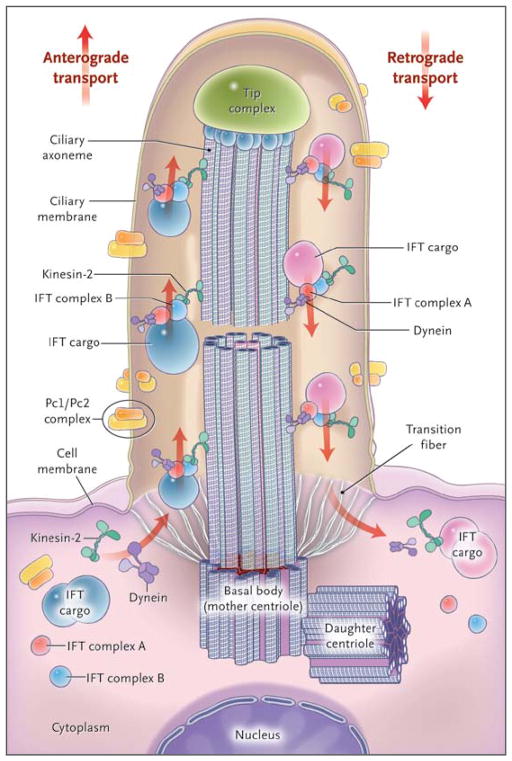
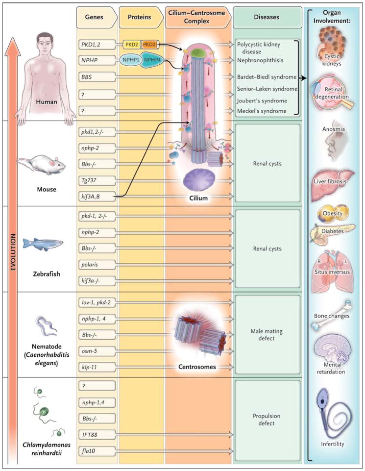
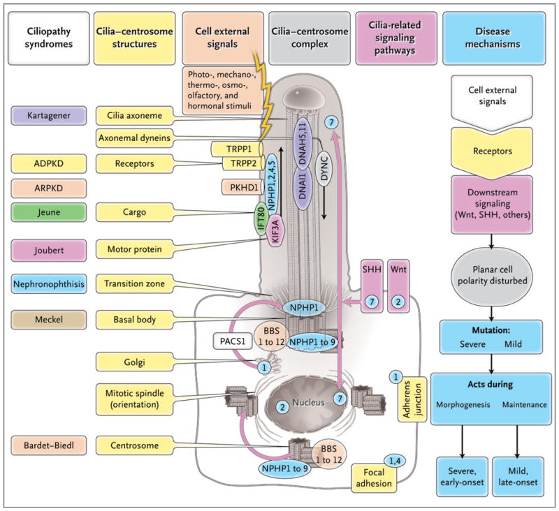
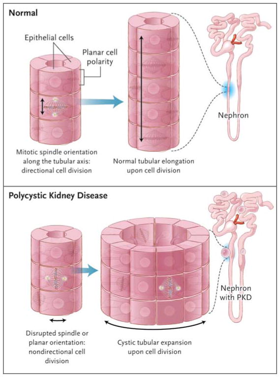
Diverse developmental and degenerative single-gene disorders such as polycystic kidney disease, nephronophthisis, retinitis pigmentosa, the Bardet–Biedl syndrome, the Joubert syndrome, and the Meckel syndrome may be categorized as ciliopathies — a recent concept that describes diseases characterized by dysfunction of a hairlike cellular organelle called the cilium. Most of the proteins that are altered in these single-gene disorders function at the level of the cilium–centrosome complex, which represents nature’s universal system for cellular detection and management of external signals. Cilia are microtubule-based structures found on almost all vertebrate cells. They originate from a basal body, a modified centrosome, which is the organelle that forms the spindle poles during mitosis. The important role that the cilium–centrosome complex plays in the normal function of most tissues appears to account for the involvement of multiple organ systems in ciliopathies. In this review, we consider the role of the cilium in disease.


Conflict of interest statement

Dr. Benzing reports receiving lecture fees from Novartis, Amgen, and Shire; and Dr. Katsanis, lecture fees from the Beijing Genomics Institute. No other potential conflict of interest relevant to this article was reported.

Figures

Figure 1
Figure 1. Structure of the Cilium and Intraflagellar Transport
The cilium is a hairlike structure on the cell surface that consists of a microtubule-based axoneme covered by a specialized plasma membrane, which is assembled from the basal body, or mother centriole. Transition fibers act as a filter for molecules passing into or out of the cilium. Nephrocystin-1 is localized at the transition zone of epithelial cells (not shown). Axonemal and membrane components are transported in raft macromolecular particles (complexes A and B) by means of intraflagellar transport (IFT) along the axonemal doublet microtubules toward the tip complex by heterotrimeric kinesin-2. Mutations of Kif3a cause renal cysts and aplasia of the cerebellar vermis in mice. Retrograde transport occurs by means of the motor protein cytoplasmic dynein. (Adapted from Bisgrove and Yost.)
Figure 2
Figure 2. Evolutionary Conservation of Ciliopathy Genes and Organ Involvement
The cilium–centrosome complex (CCC) has been conserved throughout evolution and across organ systems. The flow of genetic information is shown from left to right, from genes to proteins and their expression at the CCC and then to disease phenotypes. The vertical axis represents evolutionary time. There is strong evolutionary conservation of “ciliopathy genes.” Many ciliopathy proteins directly interact. The pathogeneses of ciliopathies converge at cilia and centrosomes. Many of the orthologues of genes in vertebrate cystic kidney disease (e.g., lov-1 or nph-4) are expressed in ciliated neurons of the head and tail of the nematode Caenorhabditis elegans, where they cause a phenotype of a male mating defect if knocked down. Many proteins altered in the Bardet–Biedl syndrome are conserved as basal body components of motile cilia of Chlamydomonas reinhardtii, in which mutations lead to a phenotype of defective intraflagellar transport or propulsion. Sensory cilia and centrosomes are key to ciliopathies because they serve distinct functions in different tissues, causing a broad range of organ involvement. (Adapted from Hildebrandt and Zhou.)
Figure 3
Figure 3. Ciliopathy Proteins and Their Relationships to the Cilium–Centrosome Complex (CCC)
Single-gene ciliopathies are shown, with colors matching the respective gene products located at the CCC machinery. Subcellular components of the CCC can be seen within a ciliated epithelial cell and include polycystin-1 (TRPP1), polycystin-2 (TRPP2), fibrocystin-polyductin (PKHD1), intraflagellar-transport (IFT) cargo, kinesin anterograde motor components (KIF3A), and cytoplasmic dynein (DYNC). Receptors on cilia perceive cell external signals and process them through the Wnt, sonic hedgehog, and focal adhesion signaling pathways. These pathways play a role in planar cell polarity, which is mediated partially through the orientation of centrosomes and the mitotic spindle poles. Depending on the severity of mutations within the same gene (e.g., in nephronophthisis type 6 [NPHP6]), they may act either during morphogenesis to cause a severe, early-onset, developmental disease phenotype (e.g., Meckel’s syndrome) or during tissue maintenance and repair to cause a mild, late-onset, degenerative disease phenotype (e.g., the Senior-Løken syndrome). The numbers in blue circles denote subcellular sites of different nephrocystins (NPHP1, 2, 4, 5, and 7).
Figure 4
Figure 4. Defects in the Noncanonical Wnt/PCP Pathway Leading to Renal Cysts
Correct orientation of the mitotic spindle and centrosomes with respect to the longitudinal axis of the tubule is critical for proper planar cell polarity (i.e., the orientation of an epithelial cell layer in three-dimensional space). Noncanonical Wnt/PCP signaling is involved in the regulation of planar cell polarity during renal tubular morphogenesis, when in 2-week-old rodents the tubules still elongate. In this model, if the mitotic spindle is maloriented, such as at an oblique angle to (or, to cite an extreme example, perpendicular to) the longitudinal orientation of tubule growth, the resulting structure would be a dilated tubule or cyst.

References

    1. Fliegauf M, Horvath J, von Schnakenburg C, et al. Nephrocystin specifically localizes to the transition zone of renal and respiratory cilia and photoreceptor connecting cilia. J Am Soc Nephrol. 2006;17:2424–33. - PubMed
    1. Kozminski KG, Johnson KA, Forscher P, Rosenbaum JL. A motility in the eukaryotic flagellum unrelated to flagellar beating. Proc Natl Acad Sci U S A. 1993;90:5519–23. - PMC - PubMed
    1. Lin F, Hiesberger T, Cordes K, et al. Kidney-specific inactivation of the KIF3A subunit of kinesin-II inhibits renal ciliogenesis and produces polycystic kidney disease. Proc Natl Acad Sci U S A. 2003;100:5286–91. - PMC - PubMed
    1. Bisgrove BW, Yost HJ. The roles of cilia in developmental disorders and disease. Development. 2006;133:4131–43. - PubMed
    1. Rosenbaum JL, Witman GB. Intraflagellar transport. Nat Rev Mol Cell Biol. 2002;3:813–25. - PubMed

Publication types

Substances