

Segmentation of liver, its vessels and lesions from CT images for surgical planning
- PMID: 21507229
- PMCID: PMC3094217
- DOI: 10.1186/1475-925X-10-30
Segmentation of liver, its vessels and lesions from CT images for surgical planning
Abstract
Background: Cancer treatments are complex and involve different actions, which include many times a surgical procedure. Medical imaging provides important information for surgical planning, and it usually demands a proper segmentation, i.e., the identification of meaningful objects, such as organs and lesions. This study proposes a methodology to segment the liver, its vessels and nodules from computer tomography images for surgical planning.
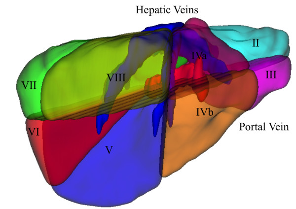
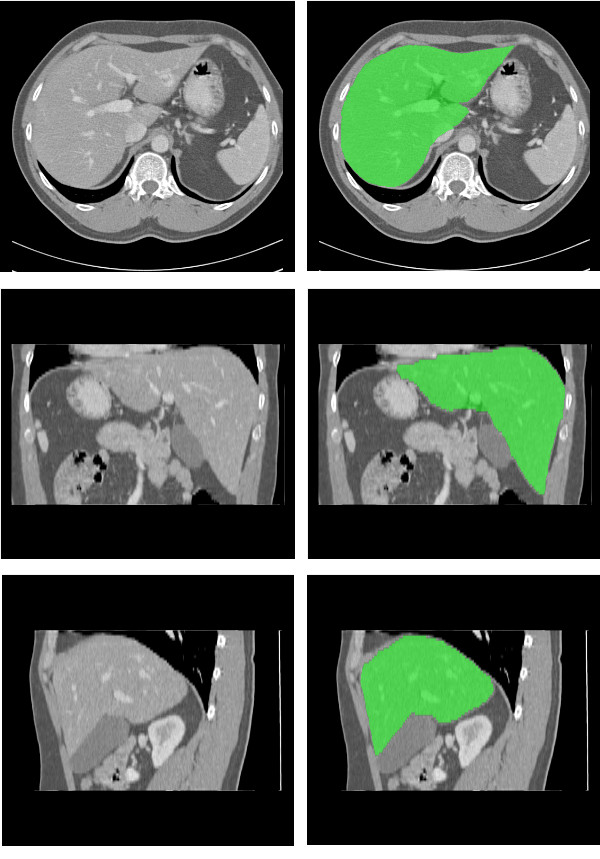
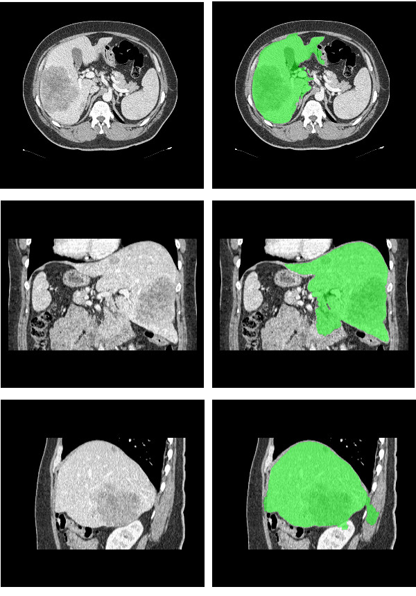
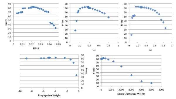
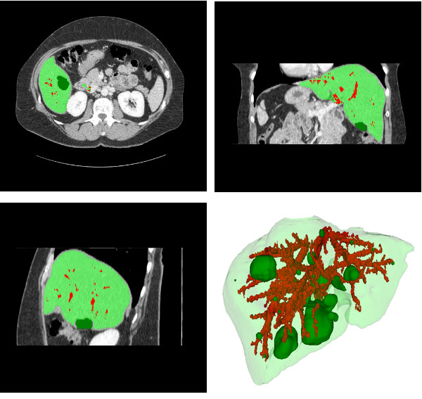
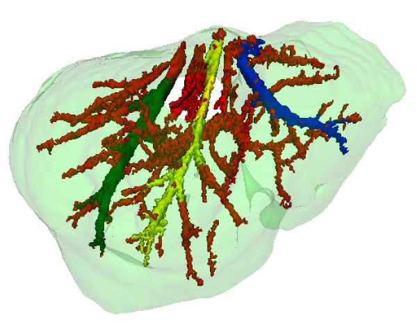
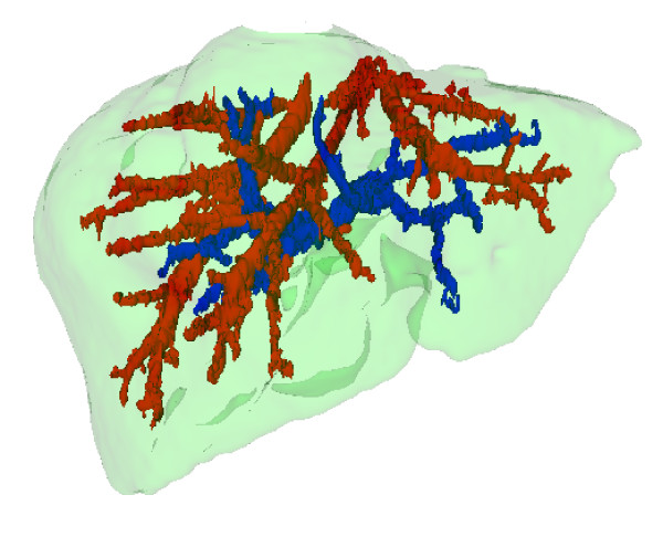
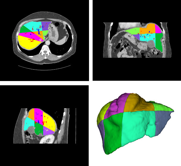
Methods: The proposed methodology consists of four steps executed sequentially: segmentation of liver, segmentation of vessels and nodules, identification of hepatic and portal veins, and segmentation of Couinaud anatomical segments. Firstly, the liver is segmented by a method based on a deformable model implemented through level sets, of which parameters are adjusted by using a supervised optimization procedure. Secondly, a mixture model is used to segment nodules and vessels through a region growing process. Then, the identification of hepatic and portal veins is performed using liver anatomical knowledge and a vein tracking algorithm. Finally, the Couinaud anatomical segments are identified according to the anatomical liver model proposed by Couinaud.
Results: Experiments were conducted using data and metrics brought from the liver segmentation competition held in the Sliver07 conference. A subset of five exams was used for estimation of segmentation parameter values, while 15 exams were used for evaluation. The method attained a good performance in 17 of the 20 exams, being ranked as the 6th best semi-automatic method when comparing to the methods described on the Sliver07 website (2008). It attained visual consistent results for nodules and veins segmentation, and we compiled the results, showing the best, worst, and mean results for all dataset.
Conclusions: The method for liver segmentation performed well, according to the results of the numerical evaluation implemented, and the segmentation of liver internal structures were consistent with the anatomy of the liver, as confirmed by a specialist. The analysis provided evidences that the method to segment the liver may be applied to segment other organs, especially to those whose distribution of voxel intensities is nearly Gaussian shaped.
Figures
Similar articles
-
Segmentation of liver and vessels from CT images and classification of liver segments for preoperative liver surgical planning in living donor liver transplantation.Comput Methods Programs Biomed. 2018 May;158:41-52. doi: 10.1016/j.cmpb.2017.12.008. Epub 2017 Dec 12. Comput Methods Programs Biomed. 2018. PMID: 29544789
-
Automated segmentation of liver and hepatic vessels on portal venous phase computed tomography images using a deep learning algorithm.J Appl Clin Med Phys. 2024 Aug;25(8):e14397. doi: 10.1002/acm2.14397. Epub 2024 May 21. J Appl Clin Med Phys. 2024. PMID: 38773719 Free PMC article.
-
Blood vessel-based liver segmentation using the portal phase of an abdominal CT dataset.Med Phys. 2013 Nov;40(11):113501. doi: 10.1118/1.4823765. Med Phys. 2013. PMID: 24320472
-
Blood vessel segmentation algorithms - Review of methods, datasets and evaluation metrics.Comput Methods Programs Biomed. 2018 May;158:71-91. doi: 10.1016/j.cmpb.2018.02.001. Epub 2018 Feb 10. Comput Methods Programs Biomed. 2018. PMID: 29544791 Review.
-
Algorithms for Liver Segmentation in Computed Tomography Scans: A Historical Perspective.Sensors (Basel). 2024 Mar 8;24(6):1752. doi: 10.3390/s24061752. Sensors (Basel). 2024. PMID: 38544015 Free PMC article. Review.
Cited by
-
Adaptive Mesh Expansion Model (AMEM) for liver segmentation from CT image.PLoS One. 2015 Mar 13;10(3):e0118064. doi: 10.1371/journal.pone.0118064. eCollection 2015. PLoS One. 2015. PMID: 25769030 Free PMC article.
-
Survey on Liver Tumour Resection Planning System: Steps, Techniques, and Parameters.J Digit Imaging. 2020 Apr;33(2):304-323. doi: 10.1007/s10278-019-00262-8. J Digit Imaging. 2020. PMID: 31428898 Free PMC article. Review.
-
Efficient Liver Segmentation from Computed Tomography Images Using Deep Learning.Comput Intell Neurosci. 2022 May 18;2022:2665283. doi: 10.1155/2022/2665283. eCollection 2022. Comput Intell Neurosci. 2022. Retraction in: Comput Intell Neurosci. 2023 Dec 13;2023:9851897. doi: 10.1155/2023/9851897. PMID: 35634046 Free PMC article. Retracted.
-
Applicability of Augmented Reality in an Organ Transplantation.Ann Transplant. 2020 Jul 31;25:e923597. doi: 10.12659/AOT.923597. Ann Transplant. 2020. PMID: 32732862 Free PMC article. Review.
-
Hepatic vessels segmentation using deep learning and preprocessing enhancement.J Appl Clin Med Phys. 2023 May;24(5):e13966. doi: 10.1002/acm2.13966. Epub 2023 Mar 18. J Appl Clin Med Phys. 2023. PMID: 36933239 Free PMC article.
References
-
- Lamecker H, Zachow S, Haberl H, Stiller M. In: Book Computer Aided Surgery around the Head. S. Weber, editor. Berlin: VDI; 2005. Medical Applications for Statistical 3D Shape Models; p. 61. vol. 17. pp. 61.
-
- Soler L, Delingette H, Malandain G, Montagnat J, Ayache N, Clement JM, Koehl C, Dourthe O, Mutter D, Marescaux J. Fully automatic anatomical, pathological, and functional segmentation from CT scans for hepatic surgery. Computer Aided Surgery. 2001;6:131–142. doi: 10.3109/10929080109145999. - DOI - PubMed
-
- Fujimoto H, Gu L, Kaneko T. Recognition of Abdominal Organs Using 3D Mathematical Morphology. System and Computers in Japan. 2002;33:75–83.
-
- Kim S, Yoo S, Kim S, Kim J, Park J. Book 5th International Conference on Signal Processing Proceedings. Vol. 2. Beijing; 2000. Segmentation of kidney without using contrast medium on abdominal CT image; pp. 1147–1152. pp. 1147-1152.
Publication types
MeSH terms
LinkOut - more resources
Full Text Sources
Other Literature Sources
Medical
