Hydroxyapatite nanoparticle-containing scaffolds for the study of breast cancer bone metastasis
- PMID: 21507478
- PMCID: PMC3613283
- DOI: 10.1016/j.biomaterials.2011.03.055
Hydroxyapatite nanoparticle-containing scaffolds for the study of breast cancer bone metastasis
Abstract
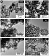
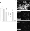
Breast cancer frequently metastasizes to bone, where it leads to secondary tumor growth, osteolytic bone degradation, and poor clinical prognosis. Hydroxyapatite Ca(10)(PO(4))(6)(OH)(2) (HA), a mineral closely related to the inorganic component of bone, may be implicated in these processes. However, it is currently unclear how the nanoscale materials properties of bone mineral, such as particle size and crystallinity, which change as a result of osteolytic bone remodeling, affect metastatic breast cancer. We have developed a two-step hydrothermal synthesis method to obtain HA nanoparticles with narrow size distributions and varying crystallinity. These nanoparticles were incorporated into gas-foamed/particulate leached poly(lactide-co-glycolide) scaffolds, which were seeded with metastatic breast cancer cells to create mineral-containing scaffolds for the study of breast cancer bone metastasis. Our results suggest that smaller, poorly-crystalline HA nanoparticles promote greater adsorption of adhesive serum proteins and enhance breast tumor cell adhesion and growth relative to larger, more crystalline nanoparticles. Conversely, the larger, more crystalline HA nanoparticles stimulate enhanced expression of the osteolytic factor interleukin-8 (IL-8). Our data suggest an important role for nanoscale HA properties in the vicious cycle of bone metastasis and indicate that mineral-containing tumor models may be excellent tools to study cancer biology and to define design parameters for non-tumorigenic mineral-containing or mineralized matrices for bone regeneration.
Copyright © 2011 Elsevier Ltd. All rights reserved.
Figures


Similar articles
-
Hydroxyapatite mineral enhances malignant potential in a tissue-engineered model of ductal carcinoma in situ (DCIS).Biomaterials. 2019 Dec;224:119489. doi: 10.1016/j.biomaterials.2019.119489. Epub 2019 Sep 11. Biomaterials. 2019. PMID: 31546097 Free PMC article.
-
Multiscale characterization of the mineral phase at skeletal sites of breast cancer metastasis.Proc Natl Acad Sci U S A. 2017 Oct 3;114(40):10542-10547. doi: 10.1073/pnas.1708161114. Epub 2017 Sep 18. Proc Natl Acad Sci U S A. 2017. PMID: 28923958 Free PMC article.
-
Tissue-engineered composite scaffold of poly(lactide-co-glycolide) and hydroxyapatite nanoparticles seeded with autologous mesenchymal stem cells for bone regeneration.J Zhejiang Univ Sci B. 2017 Nov.;18(11):963-976. doi: 10.1631/jzus.B1600412. J Zhejiang Univ Sci B. 2017. PMID: 29119734 Free PMC article.
-
Breast cancer metastasis to the bone: mechanisms of bone loss.Breast Cancer Res. 2010;12(6):215. doi: 10.1186/bcr2781. Epub 2010 Dec 16. Breast Cancer Res. 2010. PMID: 21176175 Free PMC article. Review.
-
Advances in the biology of bone metastasis: how the skeleton affects tumor behavior.Bone. 2011 Jan;48(1):6-15. doi: 10.1016/j.bone.2010.07.015. Epub 2010 Jul 17. Bone. 2011. PMID: 20643235 Review.
Cited by
-
Protein-crystal interface mediates cell adhesion and proangiogenic secretion.Biomaterials. 2017 Feb;116:174-185. doi: 10.1016/j.biomaterials.2016.11.043. Epub 2016 Nov 25. Biomaterials. 2017. PMID: 27940370 Free PMC article.
-
Exploring bone-tumor interactions through 3D in vitro models: Implications for primary and metastatic cancers.J Bone Oncol. 2025 Jun 17;53:100698. doi: 10.1016/j.jbo.2025.100698. eCollection 2025 Aug. J Bone Oncol. 2025. PMID: 40606222 Free PMC article. Review.
-
Hydroxyapatite mineral enhances malignant potential in a tissue-engineered model of ductal carcinoma in situ (DCIS).Biomaterials. 2019 Dec;224:119489. doi: 10.1016/j.biomaterials.2019.119489. Epub 2019 Sep 11. Biomaterials. 2019. PMID: 31546097 Free PMC article.
-
Intrafibrillar, bone-mimetic collagen mineralization regulates breast cancer cell adhesion and migration.Biomaterials. 2019 Apr;198:95-106. doi: 10.1016/j.biomaterials.2018.05.002. Epub 2018 May 7. Biomaterials. 2019. PMID: 29759731 Free PMC article.
-
Modelling bone metastasis in spheroids to study cancer progression and screen cisplatin efficacy.Cell Prolif. 2024 Sep;57(9):e13693. doi: 10.1111/cpr.13693. Epub 2024 Jun 20. Cell Prolif. 2024. PMID: 38899562 Free PMC article.
References
-
- Elliott JC. Structure and chemistry of the apatites and other calcium orthophosphates. Elsevier. 1994
-
- Bi X, Patil C, Morrissey C, Roudier MP, Mahadevan-Jansen A, Nyman J. Characterization of bone quality in prostate cancer bone metastases using raman spectroscopy. Proceedings of SPIE. 2010;7548:75484L.
-
- Boskey A. Bone mineral crystal size. Osteoporosis International. 2003;14:S16–20. - PubMed
-
- Balasundaram G, Sato M, Webster TJ. Using hydroxyapatite nanoparticles and decreased crystallinity to promote osteoblast adhesion similar to functionalizing with RGD. Biomaterials. 2006;27(14):2798–805. - PubMed
Publication types
MeSH terms
Substances
Grants and funding
LinkOut - more resources
Full Text Sources
Other Literature Sources
Medical

