. 2011 Aug;32(22):5241-51.
doi: 10.1016/j.biomaterials.2011.03.063. Epub 2011 Apr 20.

Effects of protein dose and delivery system on BMP-mediated bone regeneration

Affiliations

Effects of protein dose and delivery system on BMP-mediated bone regeneration

Joel D Boerckel et al. Biomaterials. 2011 Aug.

Abstract

Delivery of recombinant proteins is a proven therapeutic strategy to promote endogenous repair mechanisms and tissue regeneration. Bone morphogenetic protein-2 (rhBMP-2) has been used to promote spinal fusion and repair of challenging bone defects; however, the current clinically-used carrier, absorbable collagen sponge, requires high doses and has been associated with adverse complications. We evaluated the hypothesis that the relationship between protein dose and regenerative efficacy depends on delivery system. First, we determined the dose-response relationship for rhBMP-2 delivered to 8-mm rat bone defects in a hybrid nanofiber mesh/alginate delivery system at six doses ranging from 0 to 5 μg. Next, we directly compared the hybrid delivery system to the collagen sponge at 0.1 and 1.0 μg. Finally, we compared the in vivo protein release properties of the two delivery methods. In the hybrid delivery system, bone volume, connectivity and mechanical properties increased in a dose-dependent manner to rhBMP-2. Consistent bridging of the defect was observed for doses of 1.0 μg and greater. Compared to collagen sponge delivery at the same 1.0 μg dose, the hybrid system yielded greater connectivity by week 4 and 2.5-fold greater bone volume by week 12. These differences may be explained by the significantly greater protein retention in the hybrid system compared to collagen sponge. This study demonstrates a clear dose-dependent effect of rhBMP-2 delivered using a hybrid nanofiber mesh/alginate delivery system. Furthermore, the effective dose was found to vary with delivery system, demonstrating the importance of biomaterial carrier properties in the delivery of recombinant proteins.


Figures

Figure 1
Figure 1
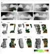
Representative digital radiographs (A) and microCT reconstructions (B) showing 3-dimensional structure and saggital cross sections illustrating local mineral density mapping. Segmental defects were treated with 0.0, 0.1, 0.5, 1.0, 2.5, and 5.0 μg rhBMP-2, as indicated, delivered in the nanofiber mesh/alginate delivery system.
Figure 2
Figure 2
A, B: MicroCT evaluation of bone volume and connectivity, respectively, as a function of rhBMP-2 dose at week 4 (light dashed lines), week 8 (bold dashed lines), and week 12 (solid lines). Bone volume (A) and connectivity (B) demonstrated nonlinear dose-dependent responses to rhBMP-2, with a reduction in response to increased dose above 1.0 μg. C, D: Postmortem biomechanical properties as a function of rhBMP-2 dose. Torsional stiffness (C) and failure torque (D) continuously increased with increasing dose of rhBMP-2. a: p < 0.05 as indicated, b: p < 0.05 vs. all other groups.
Figure 3
Figure 3
Week 12 histological staining of saggital sections at each dose of BMP-2, delivered in the mesh/alginate delivery system. H&E staining (A) illustrated bone formation (white arrow) and cellular invasion. Images at 20x, scale bars: 50 μm. Safranin-O/fast green staining at 4x (B) illustrated dose-dependent increases in alginate gel (black arrow) fragmentation and degradation as well as tissue infiltration (fast green counterstain). Images at 4x, scale bars: 200 μm.
Figure 4
Figure 4
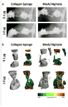
Week 12 digital radiographs (A) of segmental defects treated with 0.1 or 1.0 μg rhBMP-2, delivered in either collagen sponge or in the nanofiber mesh/alginate delivery system. MicroCT reconstructions (B) and saggital cross sections with local mineral density mapping to illustrate bone formation, defect bridging and tissue maturity.
Figure 5
Figure 5
In vivo microCT quantification of bone volume (A–C) and connectivity (D–E) at week 4 (A, D), week 8 (B, E) and week 12 (C, F) post-surgery. Dark bars represent mesh/alginate delivery system and light bars represent collagen sponge delivery system. Mesh/alginate delivery yielded an early increase in connectivity (D) and conferred a 2.5-fold greater bone volume by week 12 (C) in comparison to collagen sponge delivery. Post-mortem biomechanical testing (G, H) revealed significant dose-dependent increases in stiffness (G) and failure torque (H) but differences between delivery systems were not significant at either dose. a: p < 0.05 as indicated, c: p < 0.05 vs. same group at week 4.
Figure 6
Figure 6
Week 12 histological staining of saggital sections. H&E staining (A) illustrated bone (b) and fibrous tissue (f) formation and residual alginate (a). Images at 20x, scale bars: 50 μm. Staining with Safranin-O (B) revealed a significant persistence of alginate gel (a) through week 12 in the mesh/alginate group. Large amounts of fibrous tissue (f) were apparent in the collagen sponge group at 0.1 μg, while at 1.0 μg small amounts of trabecular bone (b) and fatty marrow filled the defect. The collagen sponge had entirely absorbed by week 12 in both collagen sponge groups. Images at 4x, scale bars: 200 μm.
Figure 7
Figure 7
In vivo tracking of fluorescent tag-labeled rhBMP-2 (inset) over 21 days revealed a significantly elevated protein retention in the mesh/alginate group compared to collagen sponge at day 3 and 7 post-implantation. Solid lines represent curve fit to exponential decay (R2 = 0.946 and 0.857 for collagen sponge and mesh/alginate groups, respectively). The half life of release was 1.87 days (95% CI: 1.49 to 2.49 days) and 3.19 days (95% CI: 2.23 to 5.59 days), for the collagen sponge and mesh/alginate, respectively.

References

    1. Sasso RC, LeHuec JC, Shaffrey C. Iliac crest bone graft donor site pain after anterior lumbar interbody fusion: a prospective patient satisfaction outcome assessment. J Spinal Disord Tech. 2005;18(Suppl):S77–81. - PubMed
    1. Rihn JA, Kirkpatrick K, Albert TJ. Graft options in posterolateral and posterior interbody lumbar fusion. Spine (Phila Pa 1976) 2010;3517:1629–1639. - PubMed
    1. Koefoed M, Ito H, Gromov K, Reynolds DG, Awad HA, Rubery PT, et al. Biological effects of rAAV-caAlk2 coating on structural allograft healing. Mol Ther. 2005;122:212–218. - PubMed
    1. Berrey BH, Jr, Lord CF, Gebhardt MC, Mankin HJ. Fractures of allografts. Frequency, treatment, and end-results. J Bone Joint Surg Am. 1990;726:825–833. - PubMed
    1. Zhang X, Awad HA, O’Keefe RJ, Guldberg RE, Schwarz EM. A perspective: engineering periosteum for structural bone graft healing. Clin Orthop Relat Res. 2008;4668:1777–1787. - PMC - PubMed

Publication types

MeSH terms