. 2011 Jun 10;286(23):20952-62.
doi: 10.1074/jbc.M111.237446. Epub 2011 Apr 20.

Mapping the intermedilysin-human CD59 receptor interface reveals a deep correspondence with the binding site on CD59 for complement binding proteins C8alpha and C9

Affiliations

Mapping the intermedilysin-human CD59 receptor interface reveals a deep correspondence with the binding site on CD59 for complement binding proteins C8alpha and C9

Stephanie E Wickham et al. J Biol Chem. .

Abstract

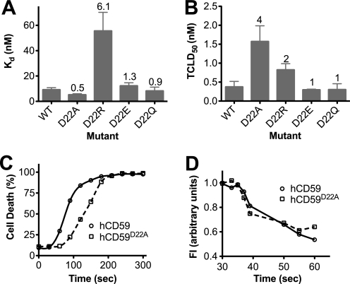
CD59 is a glycosylphosphatidylinositol-anchored protein that inhibits the assembly of the terminal complement membrane attack complex (MAC) pore, whereas Streptococcus intermedius intermedilysin (ILY), a pore forming cholesterol-dependent cytolysin (CDC), specifically binds to human CD59 (hCD59) to initiate the formation of its pore. The identification of the residues of ILY and hCD59 that form their binding interface revealed a remarkably deep correspondence between the hCD59 binding site for ILY and that for the MAC proteins C8α and C9. ILY disengages from hCD59 during the prepore to pore transition, suggesting that loss of this interaction is necessary to accommodate specific structural changes associated with this transition. Consistent with this scenario, mutants of hCD59 or ILY that increased the affinity of this interaction decreased the cytolytic activity by slowing the transition of the prepore to pore but not the assembly of the prepore oligomer. A signature motif was also identified in the hCD59 binding CDCs that revealed a new hCD59-binding member of the CDC family. Although the binding site on hCD59 for ILY, C8α, and C9 exhibits significant homology, no similarity exists in their binding sites for hCD59. Hence, ILY and the MAC proteins interact with common amino acids of hCD59 but lack detectable conservation in their binding sites for hCD59.


Figures

FIGURE 1.
FIGURE 1.
Human CD59 structure. Shown are representations of the NMR structure of the glycosylated form of the human CD59 molecule (34). Shown in a red ribbon representation is the variable region previously shown to contribute to ILY binding (10). Shown in yellow space-filled atoms are the positions of the disulfide forming cysteines and in the cyan space-filled atoms is the N-glycosylation at Asn-16. Shown in green space-filled atoms is Asn-77, which is attached to the GPI anchor. The residues examined in this study are shown in a transparent blue surface in the left figure and solid blue surface in the right figure (180° rotation). The structural representations were generated in VMD (40).
FIGURE 2.
FIGURE 2.
Effect of hCD59 mutants on ILY binding affinity and cytolytic activity. A, CHO cells transfected with hCD59 mutants were incubated with various concentrations of fluorescently modified monomer-locked ILY (ILYml) for 1 h at 37 °C, and then binding was analyzed by flow cytometry to determined the level of bound ILYml from which the Kd was determined. Monomer-locked ILY was used to minimize the affects of avidity resulting from oligomerization of the ILY monomers on the membrane. B, CHO cells transfected with hCD59 mutants were also assayed for changes in their susceptibility to ILY cell lysis by determining the TCLD50 (tissue culture lethal dose of ILY required for 50% cell death). Cells were treated with various amounts of ILYwt for 1 h at 37 °C, and cell death was determined by propidium iodide staining and flow cytometry. The numbers above the bars indicate the -fold change in Kd or TCLD50 over wild type hCD59. Shown is the average of five independent analyses. ND, not detected, neither appreciable binding (B) nor cell lysis (A) was detected above background at the concentrations used.
FIGURE 3.
FIGURE 3.
Effect of mutation of hCD59 Asp-22 on the ILY pore-forming mechanism. A, CHO cells transfected with hCD59 Asp-22 mutants were incubated with various concentrations of fluorescently labeled ILYml for 1 h at 37 °C to determine binding affinity by flow cytometry. B, CHO cells expressing the hCD59 Asp-22 substitutions were assayed for sensitivity to lysis by native ILY. The numbers above the bars indicated the -fold change in Kd or TCLD50 of ILY for each hCD59 Asp-22 mutant compared with wild type hCD59. C, the rate of cell lysis was determined for CHO cells expressing wild type hCD59 (solid lines) or hCD59D22A (dashed lines). Cells were incubated in media containing propidium iodide at 37 °C, and ILY was added to the cells at 30 s. Samples were taken and measured for PI uptake every 30 s by flow cytometry. D, the rate of oligomerization was determined by FRET between donor- and acceptor-labeled prepore-locked ILY (ILYpp). Prepore-locked ILY allowed the rate of oligomerization to be measured in the absence of cell lysis due to pore formation. Alexa488-labeled ILYpp (donor) was incubated with an equimolar amount of tetramethylrhodamine labeled ILYpp (acceptor) on CHO cells expressing wild type hCD59 (solid lines) or hCD59D22A (dashed lines). The rate of oligomerization was determined by monitoring the rate in change in donor fluorescence quenching due to FRET with acceptor-labeled ILY over time (see “Materials and Methods” for details). For C and D data are representative of 3–6 experiments. Experimental details for lytic and oligomerization assays are described in the “ILY Oligomerization Kinetics,” “Binding Affinity Analysis,“ and “Cell Lysis Assays” sections under “Materials and Methods.” FI, fluorescence intensity.
FIGURE 4.
FIGURE 4.
Residues of hCD59 necessary for ILY binding also contribute to its complement inhibitory function. CHO cells expressing the various hCD59 mutants were assayed for susceptibility to complement-mediated cell lysis. A, cells were sensitized with rabbit anti-CHO IgG antibody (26), human serum (10% v/v) was added as the source of complement components, and the cells were incubated for 45 min at 37 °C. B, as shown in Fig. 2, substitution of Asp-22 with different amino acids either increased or decreased binding affinity of ILY. The protective capacity of these mutants to MAC-mediated lysis is shown. Cell death was determined by PI staining and flow cytometry. Shown is the average of five independent analyses. Details are described under “Binding Affinity Analysis” under “Materials and Methods.”
FIGURE 5.
FIGURE 5.
ILY residues that contribute to the hCD59 binding site. A, blots of CHAPS-extracted human erythrocyte membrane proteins were probed with the ILY, ILYYY, and ILYRS (20 nm) or with the anti-CD59 monoclonal antibody H19. Each lane was loaded with the same amount of CHAPS-extracted membrane protein obtained from a single preparation of CHAPS-extracted human erythrocyte membrane proteins. B, human erythrocytes were incubated with fluorescently labeled ILYml or its derivatives, and changes in binding were determined by flow cytometry. Shown is the average of five independent analyses.
FIGURE 6.
FIGURE 6.
Monoclonal antibodies to hCD59 and prepore-locked ILY inhibit ILY-mediated lysis of human erythrocytes. Human erythrocytes were either untreated or pretreated with monoclonal antibodies to huCD59 (H19, MEM43, and 10G10) and then treated with different amounts of ILY (A), LLY (B), or PFO (C), and the extent of hemolysis was determined by the amount of hemoglobin released at each concentration of toxin. In D we determined if monomer-locked ILY or PFO could inhibit LLY-mediated hemolysis by binding to their cognate receptors hCD59 or cholesterol, respectively, and blocking LLY-mediated hemolysis. Human erythrocytes were either untreated or preincubated with 350 nm prepore-locked ILY or PFO and then treated with various concentrations of active LLY. The extent of LLY-mediated hemolysis was then determined. Shown are representative data of three or more experiments.
FIGURE 7.
FIGURE 7.
Computer model of the interaction between ILY and hCD59. The molecular model of the ILY domain 4 (cyan) and hCD59 (magenta) interaction is shown. A, ILY residues involved in binding to hCD59 are depicted as cyan-colored sticks, and the hCD59 residues involved in ILY binding are shown as magenta-colored sticks. ILY loops L1-L3 are shown in green, and the tryptophan-rich undecapeptide is colored gray. hCD59 residue Asn-77 is represented as space fill (colored by atom type; carbon atoms are magenta, nitrogen atoms are blue, oxygen atoms are red, and hydrogen atoms are white) and is the site of attachment to the GPI membrane anchor. B, a 180° rotation about the y axis from the view shown in A. ILY residues Arg-451 and Ser-452 are located on opposite sides of the β-strand, whereas Tyr-434 and Tyr-436 are located on the same side of the β-strand.

References

    1. Law R. H., Lukoyanova N., Voskoboinik I., Caradoc-Davies T. T., Baran K., Dunstone M. A., D'Angelo M. E., Orlova E. V., Coulibaly F., Verschoor S., Browne K. A., Ciccone A., Kuiper M. J., Bird P. I., Trapani J. A., Saibil H. R., Whisstock J. C. (2010) Nature 468, 447–451 - PubMed
    1. Rosado C. J., Buckle A. M., Law R. H., Butcher R. E., Kan W. T., Bird C. H., Ung K., Browne K. A., Baran K., Bashtannyk-Puhalovich T. A., Faux N. G., Wong W., Porter C. J., Pike R. N., Ellisdon A. M., Pearce M. C., Bottomley S. P., Emsley J., Smith A. I., Rossjohn J., Hartland E. L., Voskoboinik I., Trapani J. A., Bird P. I., Dunstone M. A., Whisstock J. C. (2007) Science 317, 1548–1551 - PubMed
    1. Hadders M. A., Beringer D. X., Gros P. (2007) Science 317, 1552–1554 - PubMed
    1. Slade D. J., Lovelace L. L., Chruszcz M., Minor W., Lebioda L., Sodetz J. M. (2008) J. Mol. Biol. 379, 331–342 - PMC - PubMed
    1. Shatursky O., Heuck A. P., Shepard L. A., Rossjohn J., Parker M. W., Johnson A. E., Tweten R. K. (1999) Cell 99, 293–299 - PubMed

Publication types

MeSH terms