. 2011 Jun 24;286(25):22122-30.
doi: 10.1074/jbc.M111.236257. Epub 2011 Apr 20.

Amyloid-β annular protofibrils evade fibrillar fate in Alzheimer disease brain

Affiliations

Amyloid-β annular protofibrils evade fibrillar fate in Alzheimer disease brain

Cristian A Lasagna-Reeves et al. J Biol Chem. .

Abstract

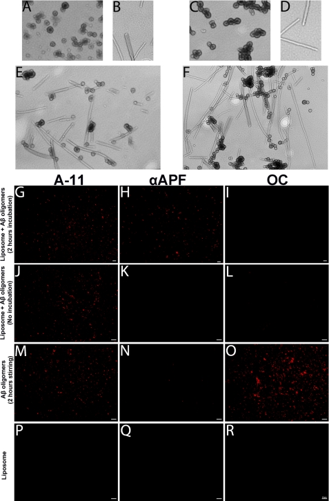
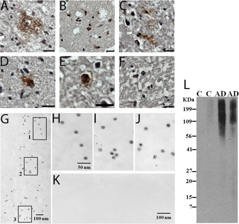
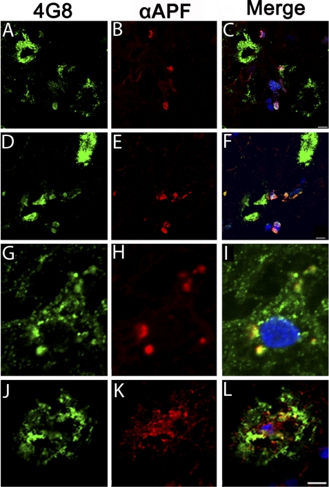
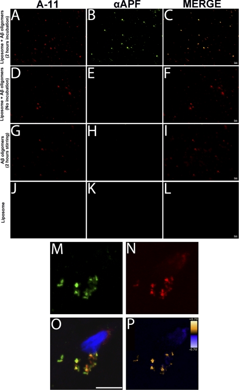
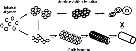
Annular protofibrils (APFs) represent a new and distinct class of amyloid structures formed by disease-associated proteins. In vitro, these pore-like structures have been implicated in membrane permeabilization and ion homeostasis via pore formation. Still, evidence for their formation and relevance in vivo is lacking. Herein, we report that APFs are in a distinct pathway from fibril formation in vitro and in vivo. In human Alzheimer disease brain samples, amyloid-β APFs were associated with diffuse plaques, but not compact plaques; moreover, we show the formation of intracellular APFs. Our results together with previous studies suggest that the prevention of amyloid-β annular protofibril formation could be a relevant target for the prevention of amyloid-β toxicity in Alzheimer disease.


Figures

FIGURE 1.
FIGURE 1.
Annular protofibrils evade fibrillar fate. A–F, pure Aβ APFs (A and C) were incubated with pure Aβ fibrils (B and D) for 48 h (E) up to 30 days (F). APFs do not convert to fibrils, as is common for oligomers, showing that APFs evade fibrillar fate. G–I, liposomes reconstituted with Aβ oligomers and incubated for 2 h exhibit punctate immunofluorescence for oligomers (A-11 column) and αAPFs but not for fibrils (OC column), suggesting that oligomers convert to APFs in the presence of membranes but not to fibrils. J–L, liposomes reconstituted with Aβ oligomers without incubation only exhibit punctate immunofluorescence for oligomers. M–O, when oligomers were stirred for 2 h without liposomes, oligomers and fibrils were detected but not APFs. P–R, as a negative control, liposomes alone were stained with A-11 and αAPF.
FIGURE 2.
FIGURE 2.
Detection of amyloid pores in tissues from Alzheimer disease brains. A–E, amyloid pores were detected in AD brain frontal cortex sections by light field microscopy after immunostaining using the 3,3′-diaminobenzidine method. The pore-like structures were present in diffuse plaques and as punctate, diffuse deposits mainly located around the nucleus, which was stained with hematoxylin. F, no amyloid pores were detected in tissues from age-matched control brains. (Scale bars represent 20 μm.) G–J, amyloid pores were isolated by immunoprecipitation using αAPF antibody and visualized by electron microscopy. Images H, I, and J are magnifications of squares 1, 2, and 3, respectively. K, no amyloid pores were pulled down by immunoprecipitation from age-matched controls. Micrographs represent immunoprecipitation from three AD cases and three control cases. L, Western blot analysis with αAPF shows that amyloid pores with molecular mass >90 kDa were present only in AD cases (AD lane). The control cases (C lane) used in this study did not present Aβ amyloid plaques.
FIGURE 3.
FIGURE 3.
Amyloid-β pores in Alzheimer disease brains were detected intracellularly and in extracellular diffuse plaques but not in compact plaques. A–F, double staining with 4G8 for Aβ (green fluorescence) and αAPF (red fluorescence) demonstrates that APFs are not associated with compact plaques and are present intracellularly. G–I, higher magnification confirmed that Aβ pores are intracellular with a punctate staining pattern characteristic of membrane vesicles. J–L, using the same immunostaining method, Aβ pores were also detected in diffuse plaques. (Scale bars represent 10 μm). Nuclei were stained with TO-PRO-3.
FIGURE 4.
FIGURE 4.
Connection between oligomers and pores in Alzheimer disease brains. A–C, liposomes reconstituted with Aβ oligomers and incubated for 2 h exhibit colocalization between punctate immunofluorescence for oligomers (A-11 column, red) and APFs (αAPF column, green), suggesting that oligomers convert to APFs. D–F, liposomes reconstituted with Aβ oligomers without incubation exhibit only punctate immunofluorescences for oligomers (A-11 column, red). G–I, the same happens with oligomers stirred for 2 h without liposomes; only oligomers were detected. J–L, as a negative control, liposomes alone were stained with A-11 and αAPF. M–O, double staining in AD brain sections with anti-oligomer specific antibody A-11 (green fluorescence) and αAPF antibody (red fluorescence) demonstrates the interactions between oligomers and pores in vivo. (Scale bar represents 10 μm.) P, the colocalization of oligomers with pores was confirmed by intensity correlation analysis of the signals. Nuclei were stained with TOP-RO-3.
FIGURE 5.
FIGURE 5.
Annular protofibrils are on a distinct pathway from amyloid fibril formation. A schematic diagram of how APFs and amyloid fibrils originate from spherical oligomers, to follow two independent amyloidogenic pathways, is shown.

Similar articles

Cited by

References

    1. Dobson C. M. (2006) Protein Pept. Lett. 13, 219–227 - PubMed
    1. Hardy J. (2005) Biochem. Soc. Trans. 33, 578–581 - PubMed
    1. Chiti F., Stefani M., Taddei N., Ramponi G., Dobson C. M. (2003) Nature 424, 805–808 - PubMed
    1. Terry R. D. (1996) J. Neuropathol. Exp. Neurol. 55, 1023–1025 - PubMed
    1. Westerman M. A., Cooper-Blacketer D., Mariash A., Kotilinek L., Kawarabayashi T., Younkin L. H., Carlson G. A., Younkin S. G., Ashe K. H. (2002) J. Neurosci. 22, 1858–1867 - PMC - PubMed

Publication types

Substances