

Multiple apoptotic defects in hematopoietic cells from mice lacking lipocalin 24p3
- PMID: 21507940
- PMCID: PMC3121516
- DOI: 10.1074/jbc.M110.216549
Multiple apoptotic defects in hematopoietic cells from mice lacking lipocalin 24p3
Abstract
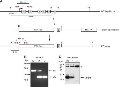
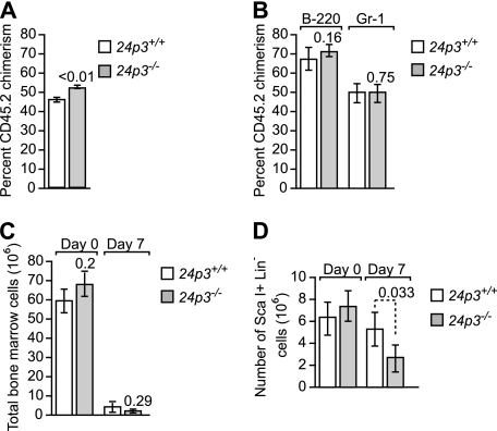
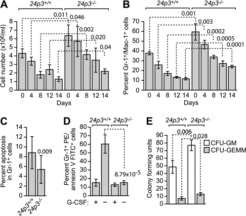
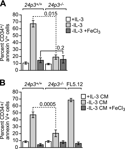
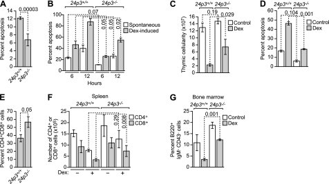
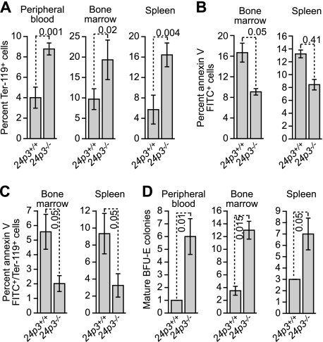
The lipocalin mouse 24p3 has been implicated in diverse physiological processes, including apoptosis, iron trafficking, development and innate immunity. Studies from our laboratory as well as others demonstrated the proapoptotic activity of 24p3 in a variety of cultured models. However, a general role for the lipocalin 24p3 in the hematopoietic system has not been tested in vivo. To study the role of 24p3, we derived 24p3 null mice and back-crossed them onto C57BL/6 and 129/SVE backgrounds. Homozygous 24p3(-/-) mice developed a progressive accumulation of lymphoid, myeloid, and erythroid cells, which was not due to enhanced hematopoiesis because competitive repopulation and recovery from myelosuppression were the same as for wild type. Instead, apoptotic defects were unique to many mature hematopoietic cell types, including neutrophils, cytokine-dependent mast cells, thymocytes, and erythroid cells. Thymocytes isolated from 24p3 null mice also displayed resistance to apoptosis-induced by dexamethasone. Bim response to various apoptotic stimuli was attenuated in 24p3(-/-) cells, thus explaining their resistance to the ensuing cell death. The results of these studies, in conjunction with those of previous studies, reveal 24p3 as a regulator of the hematopoietic compartment with important roles in normal physiology and disease progression. Interestingly, these functions are limited to relatively mature blood cell compartments.
Figures
Similar articles
-
A cell-surface receptor for lipocalin 24p3 selectively mediates apoptosis and iron uptake.Cell. 2005 Dec 29;123(7):1293-305. doi: 10.1016/j.cell.2005.10.027. Cell. 2005. PMID: 16377569
-
Bcr-Abl-mediated suppression of normal hematopoiesis in leukemia.Oncogene. 2005 May 5;24(20):3246-56. doi: 10.1038/sj.onc.1208500. Oncogene. 2005. PMID: 15735695
-
Identification of 24p3 as a direct target of Foxo3a regulated by interleukin-3 through the phosphoinositide 3-kinase/Akt pathway.J Biol Chem. 2009 Jan 23;284(4):2187-93. doi: 10.1074/jbc.M806131200. Epub 2008 Dec 4. J Biol Chem. 2009. PMID: 19056725 Free PMC article.
-
24p3 and its receptor: dawn of a new iron age?Cell. 2005 Dec 29;123(7):1175-7. doi: 10.1016/j.cell.2005.12.008. Cell. 2005. PMID: 16377555 Review.
-
Adipocytokines and insulin resistance: the possible role of lipocalin-2, retinol binding protein-4, and adiponectin.Diabetes Care. 2009 Nov;32 Suppl 2(Suppl 2):S362-7. doi: 10.2337/dc09-S340. Diabetes Care. 2009. PMID: 19875582 Free PMC article. Review. No abstract available.
Cited by
-
Skeletal Lipocalin-2 Is Associated with Iron-Related Oxidative Stress in ob/ob Mice with Sarcopenia.Antioxidants (Basel). 2021 May 11;10(5):758. doi: 10.3390/antiox10050758. Antioxidants (Basel). 2021. PMID: 34064680 Free PMC article.
-
Mammalian siderophores, siderophore-binding lipocalins, and the labile iron pool.J Biol Chem. 2012 Apr 20;287(17):13524-31. doi: 10.1074/jbc.R111.311829. Epub 2012 Mar 2. J Biol Chem. 2012. PMID: 22389496 Free PMC article. Review.
-
Lipocalin 24p3 Induction in Colitis Adversely Affects Inflammation and Contributes to Mortality.Front Immunol. 2019 Apr 17;10:812. doi: 10.3389/fimmu.2019.00812. eCollection 2019. Front Immunol. 2019. PMID: 31057545 Free PMC article.
-
Lipocalin-2 (Lcn2) expression is mediated by maternal nutrition during the development of the fetal liver.Genes Nutr. 2014 Jan;9(1):380. doi: 10.1007/s12263-013-0380-4. Epub 2014 Jan 3. Genes Nutr. 2014. PMID: 24382649 Free PMC article.
-
The Lipocalin2 Gene is Regulated in Mammary Epithelial Cells by NFκB and C/EBP In Response to Mycoplasma.Sci Rep. 2020 May 6;10(1):7641. doi: 10.1038/s41598-020-63393-x. Sci Rep. 2020. PMID: 32376831 Free PMC article.
References
-
- Akerstrom B., Flower D. R., Salier J. P. (2000) Biochim. Biophys. Acta 1482, 1–8 - PubMed
-
- Devireddy L. R., Gazin C., Zhu X., Green M. R. (2005) Cell 123, 1293–1305 - PubMed
-
- Devireddy L. R., Teodoro J. G., Richard F. A., Green M. R. (2001) Science 293, 829–834 - PubMed
-
- Dong X., Mo Z., Bokoch G., Guo C., Li Z., Wu D. (2005) Curr. Biol. 15, 1874–1879 - PubMed
Publication types
MeSH terms
Substances
Grants and funding
LinkOut - more resources
Full Text Sources
Other Literature Sources
Medical
Molecular Biology Databases
