. 2011 Jun 10;286(23):20930-41.
doi: 10.1074/jbc.M111.228213. Epub 2011 Apr 19.

The Leishmania donovani UMP synthase is essential for promastigote viability and has an unusual tetrameric structure that exhibits substrate-controlled oligomerization

Affiliations

The Leishmania donovani UMP synthase is essential for promastigote viability and has an unusual tetrameric structure that exhibits substrate-controlled oligomerization

Jarrod B French et al. J Biol Chem. .

Abstract

The final two steps of de novo uridine 5'-monophosphate (UMP) biosynthesis are catalyzed by orotate phosphoribosyltransferase (OPRT) and orotidine 5'-monophosphate decarboxylase (OMPDC). In most prokaryotes and simple eukaryotes these two enzymes are encoded by separate genes, whereas in mammals they are expressed as a bifunctional gene product called UMP synthase (UMPS), with OPRT at the N terminus and OMPDC at the C terminus. Leishmania and some closely related organisms also express a bifunctional enzyme for these two steps, but the domain order is reversed relative to mammalian UMPS. In this work we demonstrate that L. donovani UMPS (LdUMPS) is an essential enzyme in promastigotes and that it is sequestered in the parasite glycosome. We also present the crystal structure of the LdUMPS in complex with its product, UMP. This structure reveals an unusual tetramer with two head to head and two tail to tail interactions, resulting in two dimeric OMPDC and two dimeric OPRT functional domains. In addition, we provide structural and biochemical evidence that oligomerization of LdUMPS is controlled by product binding at the OPRT active site. We propose a model for the assembly of the catalytically relevant LdUMPS tetramer and discuss the implications for the structure of mammalian UMPS.


Figures

FIGURE 1.
FIGURE 1.
Pyrimidine biosynthesis. A, de novo biosynthetic pathway for pyrimidine biosynthesis. B, organization of genes involved in pyrimidine biosynthesis in humans and Leishmania spp. The genes encoding the enzymes responsible for the first three enzymatic steps (carbamoylphosphate synthetase, aspartate carbamoyltransferase, and dihydroorotase) are shown in blue, the gene for dihydroorotate dehydrogenase is shown in green, and the genes encoding the enzymes responsible for the final two steps (OMPDC and OPRT) are shown in red.
FIGURE 2.
FIGURE 2.
Structure of LdOMPDC. A, ribbon diagram of the structure of monofunctional LdOMPDC with sulfate molecules (space filling) in the active site. N and C mark the N and C termini of the protein. Helices are shown colored red, whereas β-strands are shown colored yellow. B, comparison of LdOMPDC dimer (green and red/yellow) to the human OMPDC dimer (blue and red/yellow).
FIGURE 3.
FIGURE 3.
The OMPDC domain of LdUMPS. A, ribbon diagram of the OMPDC domain of LdUMPS showing UMP bound in the active sites. The electron density shown is from an FoFc map calculated before adding the ligand and contoured at 2.5 σ. B, stereodiagram of the active site of the OMPDC domain of LdUMPS. The carbon atoms of residues from different OMPDC chains are colored yellow and cyan, respectively, and the UMP molecule is shown with green carbon atoms. In both cases, oxygen atoms are colored red and nitrogen atoms are colored blue.
FIGURE 4.
FIGURE 4.
Structure of LdUMPS. A, ribbon diagram of the LdUMPS protomer with the two domains labeled. Helices are shown colored red, whereas β-strands are shown colored yellow. B, LdUMPS tetramer with the four chains shown in red, yellow, green, and blue and the OPRT and OMPDC dimers highlighted by green and blue surface representation, respectively. The tetramer was constructed with two copies of the asymmetric unit related by a 2-fold rotation. The yellow/red pair and the blue/green pair each represent a single copy of the asymmetric unit.
FIGURE 5.
FIGURE 5.
Ligand-dependent oligomerization of LdUMPS. A, size exclusion chromatography of LdUMPS run in buffer without added ligand (red solid line), with 5 mm UMP added (blue dashed line), and with 500 μm OMP added (black dotted line). A UMP peak appears in all chromatograms as a result of UMP that is present in the purification of LdUMPS. B, CD spectra of LdUMPS without added UMP (black dashed line) and at two time points after adding UMP (red and blue solid lines).
FIGURE 6.
FIGURE 6.
The OPRT domain of LdUMPS. A, superposition of apo and ligand-bound S. cerevisiae OPRT (17) and the two OPRT domains from the structure of LdUMPS. B and C, active site of OPRT from the two different chains found in the asymmetric unit of the structure of LdUMPS. UMP is modeled into the density for B, and shown in a similar location in C. The electron density shown is from an FoFc map calculated before adding the ligand and contoured at 2.5 σ. D, a superposition of the two different OPRT chains shows the residues believed to be responsible for closing the hood region about the active site. The closed conformation of the enzyme is shown in green, whereas the open conformation is shown in blue, and UMP is shown colored gray. Lys279 is not shown for the chain in the open conformation as it is not visible in the LdUMPS structure.
FIGURE 7.
FIGURE 7.
Model for assembly of the biologically active LdUMPS tetramer. After expression, the bifunctional LdUMPS forms a tight dimer at the OMPDC domain. Ligand binding causes a conformational change in the OPRT domain (shown in red), which facilitates the dimerization at the OPRT domain, leading to the observed tetrameric form.
FIGURE 8.
FIGURE 8.
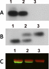
Southern and Western blot analysis of Δumps parasites. A, genomic DNA prepared from wild-type, LdUMPS/umps, and Δumps parasites was digested with PvuII and hybridized to a 1.37-kb probe from the LdUMPS ORF and B, to a 0.73-kb probe from the 5′-flanking region of LdUMPS. C, a Western blot of wild-type, LdUMPS/umps, and Δumps cell lysates fractionated on an SDS-polyacrylamide gel and probed with monospecific antisera raised against the purified LdUMPS protein and monoclonal antibody directed against mouse α-tubulin (24). Lanes 1–3 in all panels refer to DNA or protein obtained from wild-type, LdUMPS/umps, and Δumps cells, respectively.
FIGURE 9.
FIGURE 9.
Growth of Δumps promastigotes in various pyrimidine sources. The ability of wild-type (black bars) and Δumps (hashed bars) parasites to grow in medium containing orotic acid or a pyrimidine supplement was ascertained as described under “Experimental Procedures.” All supplements were present at a concentration of 100 μm, except where indicated. Data are plotted in histogram form as a percentage of maximal growth versus the supplement added to the normal culture medium. No pyr designates no pyrimidine added, and Oro2 mm indicates that orotic acid was added to the parasites at a final concentration of 2 mm. Each result provided is the mean ± S.D. of quadruplicate determinations.
FIGURE 10.
FIGURE 10.
Subcellular localization of LdUMPS. Exponentially growing L. donovani promastigotes were fixed and processed as described under “Experimental Procedures” prior to incubation with rabbit anti-LdUMPS and guinea pig anti-IMPDH antibodies. Primary antibodies were visualized at 488 nm with (A) Oregon Green-conjugated goat anti-rabbit IgG or (B) at 594 nm with rhodamine red-conjugated goat anti-guinea pig IgG secondary antibodies. C, the images in panels A and B were merged to demonstrate colocalization of LdUMPS and IMPDH. Confocal images were acquired on a Zeiss Axiovert 200M inverted microscope. The green signal is LdUMPS, whereas the red signal shows the localization of LdIMPDH.

References

    1. Connolly G. P., Duley J. A. (1999) Trends Pharmacol. Sci. 20, 218–225 - PubMed
    1. Evans D. R., Guy H. I. (2004) J. Biol. Chem. 279, 33035–33038 - PubMed
    1. Jones M. E. (1980) Annu. Rev. Biochem. 49, 253–279 - PubMed
    1. Carter N. S., Rager N., Ullman B. (2003) in Molecular and Medical Parasitology (Marr J. J., Nilsen T., Komuniecki R. eds) pp. 197–223, Academic Press Limited, London
    1. Weber G. (2001) Biochemistry 66, 1164–1173 - PubMed

Publication types

MeSH terms

Substances

LinkOut - more resources