. 2011 Jun 10;286(23):20769-77.
doi: 10.1074/jbc.M111.226605. Epub 2011 Apr 20.

Polo-like kinase1 is required for recruitment of dynein to kinetochores during mitosis

Affiliations

Polo-like kinase1 is required for recruitment of dynein to kinetochores during mitosis

Jason R Bader et al. J Biol Chem. .

Abstract

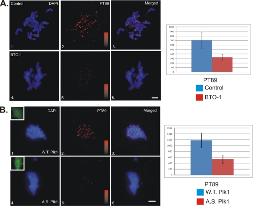
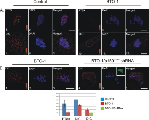
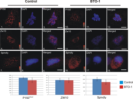
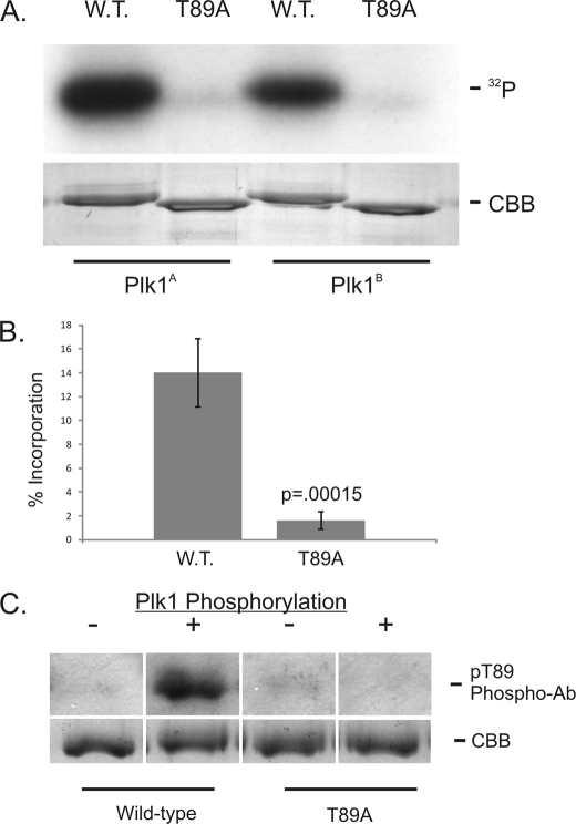
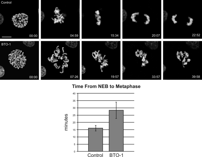
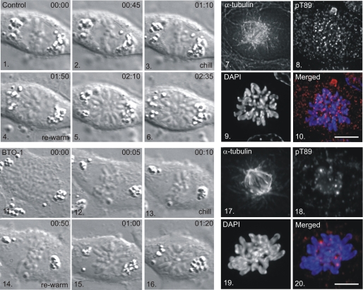
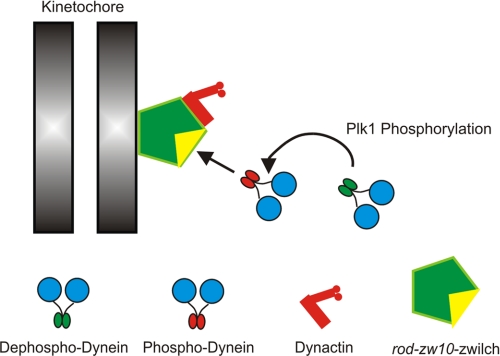
Kinetochore dynein has been implicated in microtubule capture, correcting inappropriate microtubule attachments, chromosome movement, and checkpoint silencing. It remains unclear how dynein coordinates this diverse set of functions. Phosphorylation is responsible for some dynein heterogeneity (Whyte, J., Bader, J. R., Tauhata, S. B., Raycroft, M., Hornick, J., Pfister, K. K., Lane, W. S., Chan, G. K., Hinchcliffe, E. H., Vaughan, P. S., and Vaughan, K. T. (2008) J. Cell Biol. 183, 819-834), and phosphorylated and dephosphorylated forms of dynein coexist at prometaphase kinetochores. In this study, we measured the impact of inhibiting polo-like kinase 1 (Plk1) on both dynein populations. Phosphorylated dynein was ablated at kinetochores after inhibiting Plk1 with a small molecule inhibitor (5-Cyano-7-nitro-2-(benzothiazolo-N-oxide)-carboxamide) or chemical genetic approaches. The total complement of kinetochore dynein was also reduced but not eliminated, reflecting the presence of some dephosphorylated dynein after Plk1 inhibition. Although Plk1 inhibition had a profound effect on dynein, kinetochore populations of dynactin, spindly, and zw10 were not reduced. Plk1-independent dynein was reduced after p150(Glued) depletion, consistent with the binding of dephosphorylated dynein to dynactin. Plk1 phosphorylated dynein intermediate chains at Thr-89 in vitro and generated the phospho-Thr-89 phospho-epitope on recombinant dynein intermediate chains. Finally, inhibition of Plk1 induced defects in microtubule capture and persistent microtubule attachment, suggesting a role for phosphorylated dynein in these functions during prometaphase. These findings suggest that Plk1 is a dynein kinase required for recruitment of phosphorylated dynein to kinetochores.


Figures

FIGURE 1.
FIGURE 1.
Impact of Plk1 Inhibition of Recruitment of Phospho-dynein to Kinetochores. A, NRK2 cells were treated with vehicle alone (panels 1–3) or BTO-1 (panels 4–6) and stained for chromatin (blue) and pT89 dynein (red). BTO-1 treatment resulted in a dramatic reduction in both pT89 dynein and BubR1 at kinetochores during prometaphase. Intensity scales = 0–1000 (panels 2 and 5). Scale bar = 5 μm. Statistical analysis reveals a significant difference for pT89 (p = 1.28 × 10⁻¹⁶). B, NRK2 cells were transfected with WT Plk1-GFP (panels 1–3) or AS Plk1-GFP (panels 4–6), treated with 3MB-PP1, and stained for chromatin (blue) and pT89 dynein (red). 3MB-PP1 treatment of AS but not WT Plk1 resulted in a dramatic reduction in phospho-dynein at kinetochores during prometaphase. Intensity scales = 0–1600 (panels 2 and 5). Scale bar = 5 μm. Insets display expression of GFP constructs (panels 1 and 4). Statistical analysis reveals a significant difference for pT89 (p = 5.49 × 10⁻²⁹).
FIGURE 2.
FIGURE 2.
Plk1-dependent and Independent Populations of Dynein at Kinetochores. A, NRK2 cells treated with vehicle control (panels 1–6) or BTO-1 (panels 7–12) were stained for phosphorylated dynein (PT89) or total dynein (red) and chromatin (blue). Both PT89 dynein and total dynein were reduced significantly at kinetochores after Plk1 inhibition (p = 5.16 × 10⁻²⁰ for pT89, p = 5.86 × 10⁻¹⁰ for total dynein). However, some total dynein staining remained after BTO-1 treatment. Intensity scales = 0–2500 (panels 1 and 7) and 0–3000 for total dynein (panels 4 and 10). Scale bar = 5 μm. B, control NRK2 cells and NRK2 cells expressing shRNA plasmid against p150Glued were both treated with BTO-1 and stained for total dynein (red) and chromatin (blue). Although control cells retain some total dynein after BTO-1 treatment (panels 1–3), dynein levels are reduced (p = 2.26 × 10⁻⁶) to background in cells depleted of p150Glued and treated with BTO-1 (panels 4–6). Intensity scales = 0–1500 (panels 1 and 4). Scale bar = 5 μm.
FIGURE 3.
FIGURE 3.
Impact of Plk1 Inhibition on Dynein-associated Proteins. HeLa cells treated with vehicle control (panels 1–9) or BTO-1 (panels 10–18) were stained for chromatin (blue) and p150Glued (panels 1–3 and 10–12 (red)), Zw10 (panels 4–6 and 13–15 (red)), or spindly (panels 7–9 and 16–18 (red)). None of these proteins were affected significantly by Plk1 inhibition (p > 0.05 for all three). Intensity scales = 0–4095 (panels 1 and 10), 0–250 (panels 4 and 13) or 0–2500 (panels 7 and 16). Scale bar = 5 μm.
FIGURE 4.
FIGURE 4.
In Vitro Phosphorylation of Dynein ICs by Plk1. A, Wild-type and T89A recombinant IC-2C (amino acids 1–284) were subjected to in vitro phosphorylation reactions with γ32P-ATP and purified Plk1 (Plk1A, Cell Signaling Technology; Plk1B, Cedarlane, Inc.). Equal protein loading was confirmed by Coomassie brilliant blue staining of the gel. B, Phosphorylation efficiency was measured using the specific activity of γ32P-ATP and IC protein concentration. The p value was calculated from four experiments. C, wild-type and T89A recombinant IC-2C (amino acids 1–284) were probed by Western blot analysis with the pT89 phospho-antibody before (−) or after (+) Plk1 phosphorylation in vitro. Equal loading was confirmed by Coomassie brilliant blue staining of gels run in parallel.
FIGURE 5.
FIGURE 5.
Impact of Plk1 Inhibition on Chromosome Alignment. NRK2 cells expressing mCherry-H2B were subjected to live-cell imaging after treatment with vehicle control (Control) or BTO-1. To overcome the effects of Plk1 inhibition during prophase, cells were treated at NEB and then imaged at 30-s intervals until the metaphase/anaphase transition. Control cells achieved alignment in ∼15 min after NEB, whereas treated cells failed to achieve alignment within 30 min after treatment (p < 0.05). The most common phenotype is inefficient progression to chromosome alignment (see supplemental videos SV1 and SV2). Scale bar = 5 μm.
FIGURE 6.
FIGURE 6.
Recruitment of PT89 Dynein to Kinetochores after Metaphase Alignment. NRK2 cells were monitored by differential interference contrast microscopy until chromosome alignment was achieved (panels 3 and 13) and chilled in the presence of a vehicle control (panels 1–10) or BTO-1 (panels 11–20). Cells were rewarmed (panels 4–6 and 14–16), fixed, and stained for α-tubulin (panels 7 and 17), pT89 dynein (panels 8 and 18), and chromatin (panels 9 and 19). Merged images reveal accumulation of pT89 dynein on control (panel 10) but not BTO-1-treated (panel 20) kinetochores. Scale bar = 5 μm.
FIGURE 7.
FIGURE 7.
New Model for Recruitment of Dynein to Kinetochores. Previous work suggested that phosphorylated dynein was recruited to kinetochores through a direct interaction with the rod-zw10-zwilch complex (1). However, the requirement for phosphorylation was difficult to test without kinase candidates. This study identifies Plk1 as a mitotic dynein kinase that is required for recruitment of phospho-dynein. This study provides a novel method to separate the contributions of dynactin from those of dynein alone. It also has implications for the application of Plk1 inhibition as an anti-cancer therapy.

References

    1. Whyte J., Bader J. R., Tauhata S. B., Raycroft M., Hornick J., Pfister K. K., Lane W. S., Chan G. K., Hinchcliffe E. H., Vaughan P. S., Vaughan K. T. (2008) J. Cell Biol. 183, 819–834 - PMC - PubMed
    1. Echeverri C. J., Paschal B.M., Vaughan K. T., Vallee R. B. (1996) Molecular characterization of the 50-kD subunit of dynactin reveals function for the complex in chromosome alignment and spindle organization during mitosis. J. Cell Biol. 132, 617–633 - PMC - PubMed
    1. Kops G. J., Kim Y., Weaver B. A., Mao Y., McLeod I., Yates J. R., 3rd, Tagaya M., Cleveland D. W. (2005) J. Cell Biol. 169, 49–60 - PMC - PubMed
    1. Griffis E. R., Stuurman N., Vale R. D. (2007) J. Cell Biol. 177, 1005–1015 - PMC - PubMed
    1. Gassmann R., Essex A., Hu J. S., Maddox P. S., Motegi F., Sugimoto A., O'Rourke S. M., Bowerman B., McLeod I., Yates J. R., 3rd, Oegema K., Cheeseman I. M., Desai A. (2008) Genes Dev. 22, 2385–2399 - PMC - PubMed

Publication types

MeSH terms

LinkOut - more resources