. 2011 Jun 3;286(22):20043-53.
doi: 10.1074/jbc.M111.218727. Epub 2011 Apr 20.

A disease-associated polymorphism alters splicing of the human CD45 phosphatase gene by disrupting combinatorial repression by heterogeneous nuclear ribonucleoproteins (hnRNPs)

Affiliations

A disease-associated polymorphism alters splicing of the human CD45 phosphatase gene by disrupting combinatorial repression by heterogeneous nuclear ribonucleoproteins (hnRNPs)

Laura B Motta-Mena et al. J Biol Chem. .

Abstract

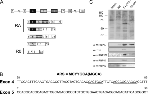
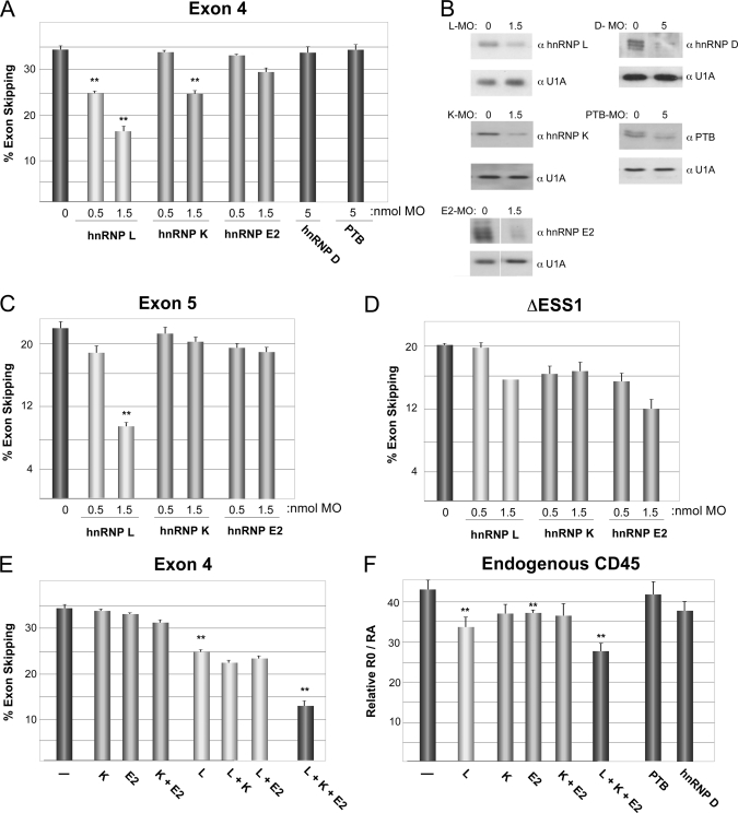
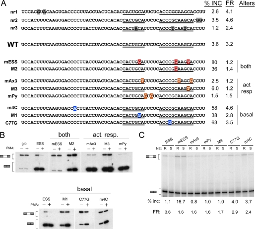
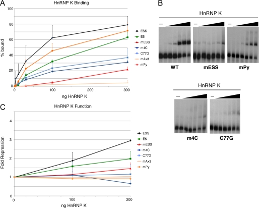
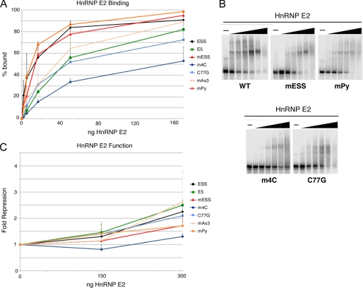
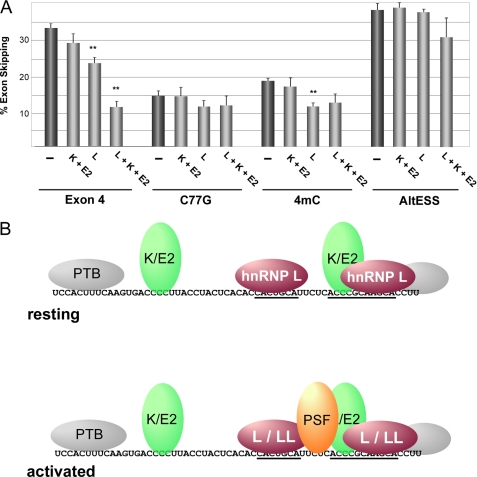
Alternative splicing is typically controlled by complexes of regulatory proteins that bind to sequences within or flanking variable exons. The identification of regulatory sequence motifs and the characterization of sequence motifs bound by splicing regulatory proteins have been essential to predicting splicing regulation. The activation-responsive sequence (ARS) motif has previously been identified in several exons that undergo changes in splicing upon T cell activation. hnRNP L binds to this ARS motif and regulates ARS-containing exons; however, hnRNP L does not function alone. Interestingly, the proteins that bind together with hnRNP L differ for different exons that contain the ARS core motif. Here we undertake a systematic mutational analysis of the best characterized context of the ARS motif, namely the ESS1 sequence from CD45 exon 4, to understand the determinants of binding specificity among the components of the ESS1 regulatory complex and the relationship between protein binding and function. We demonstrate that different mutations within the ARS motif affect specific aspects of regulatory function and disrupt the binding of distinct proteins. Most notably, we demonstrate that the C77G polymorphism, which correlates with autoimmune disease susceptibility in humans, disrupts exon silencing by preventing the redundant activity of hnRNPs K and E2 to compensate for the weakened function of hnRNP L. Therefore, these studies provide an important example of the functional relevance of combinatorial function in splicing regulation and suggest that additional polymorphisms may similarly disrupt function of the ESS1 silencer.


Figures

FIGURE 1.
FIGURE 1.
CD45 exons 4 and 5 share sequence similarity but recruit distinct proteins. A, schematic of CD45 alternative splicing, showing variable exons 4, 5, and 6 in the genomic context and the five isoforms that have been identified in human T cells. Based on antibody reactivity, isoforms encoded by mRNAs containing exon 4 are also termed RA, whereas the smallest isoform is RO. Boxes correspond to exons, and lines designate introns. B, sequence of the silencer regions of CD45 exons 4 and 5, as defined previously (Rothrock et al. (15) and Motta-Mena et al. (19)), with the ARS core element underlined and the ARS motif consensus shown above. Numbers indicate nucleotide position with respect to full exon. C, silver stain (top) and Western blot (bottom) of proteins isolated by RNA affinity using the exons 4 and 5 silencer elements (as shown in panel B), unrelated silencer sequence (NS; Melton et al. (21)), or beads alone control.
FIGURE 2.
FIGURE 2.
hnRNPs L and K have the majority of the silencing activity among the ESS1-associated proteins. A, the percentage of skipping of wild-type exon 4 in JSL1 cells upon individual knockdown of hnRNPs L, K, E, D, and PTB by transfection of the indicated amount of morpholino oligonucleotide. The average of multiple independent experiments is graphed. Corresponding S.D. and p values are shown in supplemental Table 1. Double asterisks indicate p value of less than 0.01. B, Western blots demonstrating knockdown from representative experiment used for graph in A. MO, morpholino oligonucleotide. C, the percentage of skipping of exon 5 in JSL1 cells. D, the percentage of skipping of exon 4 derivative lacking ESS1 done as for panel A. E, the percentage of skipping of exon 4 done as for panel A with 0.5 nmol of morpholino against each of the specified proteins alone or in combination. F, quantification of splicing of the endogenous CD45 gene in cells treated as in panel E. Statistics are shown in supplemental Table 1. The corresponding Western blot for panels E and F and a representative gel for panel F are shown in supplemental Fig. 1.
FIGURE 3.
FIGURE 3.
Mutation of sequences in ESS1 uncouples basal and activation-induced silencing. A, schematic of mutations used, grouped according to activity. Names of mutations are described under “Results”; nr = no response. The percentage of inclusion of mutant-containing exon in resting cells (% inc) and -fold repression difference between resting and stimulated cells (FR) is shown as an average of multiple experiments, as shown in panel B. FR is calculated as (%Exon exclusion/%Exon inclusion)activated/(%Exon exclusion/%Exon inclusion)resting. Mutations are categorized based on whether they primarily alter basal silencing (basal), the -fold repression in response to activation (act resp), or both. B, representative RT-PCR from JSL1 cells expressing the indicated minigenes grown under resting (phorbol 12-myristate 13-acetate-negative (−PMA)) or stimulated (phorbol 12-myristate 13-acetate-positive (+PMA)) conditions for 70 h. Quantification of splicing is shown in panel A. C, RT-PCR analysis of indicated minigenes spliced in vitro in nuclear extract (NE) prepared from cells grown under resting (R) or stimulated (S) conditions. The percentage of inclusion of exon 4 derivative in resting nuclear extract and -fold repression between resting and stimulated nuclear extract is shown. Statistics are shown in supplemental Table 2. In most cases, the values that deviate from ESS have a p value < 0.02.
FIGURE 4.
FIGURE 4.
Specificity of hnRNP L binding and activity. A, quantification of gel shift assays performed with purified recombinant hnRNP L and purified RNAs as indicated. B, representative gel shifts used for graph in panel A. For all panels, titration of protein is at 1, 3, 10, and 30 ng of hnRNP L per reaction. C, quantification of in vitro splicing assays in which purified recombinant hnRNP L was added to nuclear extract derived from resting cells and then incubated with minigene RNAs in which the middle exon contained wild-type exon 4, wild-type exon 5, or mutants of exon 4 as in Fig. 3. S.D. and p values are shown in supplemental Table 3.
FIGURE 5.
FIGURE 5.
Specificity of hnRNP K binding and activity. A, quantification of gel shift assays performed with purified recombinant hnRNP K and purified RNAs as indicated. B, representative gel shifts used for graph in panel A. For all panels, titration of protein is at 1, 3, 10, 30, 100, and 300 ng of hnRNP K per reaction. C, quantification of in vitro splicing assays in which purified recombinant hnRNP K was added to nuclear extract derived from resting cells and then incubated with minigene RNAs in which the middle exon contained wild-type exon 4, wild-type exon 5, or mutants of exon 4 as in Fig. 3. S.D. and p values are shown in supplemental Table 3.
FIGURE 6.
FIGURE 6.
Specificity of hnRNP E2 binding and activity. A, quantification of gel shift assays performed with purified recombinant hnRNP E2 and purified RNAs as indicated. B, representative gel shifts used for graph in panel A. For all panels, titration of protein is at 0.56, 1.7, 5.6, 17, 56, and 170 ng of hnRNP E2 per reaction. C, quantification of in vitro splicing assays in which purified recombinant hnRNP E2 was added to nuclear extract derived from resting cells and then incubated with minigene RNAs in which the middle exon contained wild-type exon 4, wild-type exon 5, or mutants of exon 4 as in Fig. 3. S.D. and p values are shown in supplemental Table 3.
FIGURE 7.
FIGURE 7.
Presence of binding site for hnRNPs K and E2 is required for their ability to compensate for reduced hnRNP L function. A, the percentage of skipping of exon 4 variants containing the wild-type ESS1 (Exon 4), ESS1 with C77G mutation (C77G), or 4mC mutation (4mC) or substitution of the ESS1 with an unrelated silencer sequence (AltESS) in JSL1 cells upon knockdown of combinations of hnRNPs L, K, and E2. Experiments were performed and analyzed as described in the legend for Fig. 2. S.D. and p values are shown in supplemental Table 4. Double asterisks indicate p < 0.01. B, model for protein association with ESS1 in resting and stimulated T cells. The core ARS motifs are underlined. Colored proteins are those shown to be functional (hnRNPs L, K, and E2 in resting cells along with hnRNP LL and PSF in activated cells). PTB is added for completeness but is in gray to indicate its lack of contribution to ESS1-dependent silencing. Proteins shown to bind to the same or immediately overlapping sites are shown as a single oval (K/E2, L/LL) for simplicity.

Similar articles

Cited by

References

    1. Wahl M. C., Will C. L., Lührmann R. (2009) Cell 136, 701–718 - PubMed
    1. Chen M., Manley J. L. (2009) Nat. Rev. Mol. Cell Biol. 10, 741–754 - PMC - PubMed
    1. Nilsen T. W., Graveley B. R. (2010) Nature 463, 457–463 - PMC - PubMed
    1. Wang E. T., Sandberg R., Luo S., Khrebtukova I., Zhang L., Mayr C., Kingsmore S. F., Schroth G. P., Burge C. B. (2008) Nature 456, 470–476 - PMC - PubMed
    1. Pan Q., Shai O., Lee L. J., Frey B. J., Blencowe B. J. (2008) Nat. Genet. 40, 1413–1415 - PubMed

Publication types

MeSH terms

Substances