. 2011 Jul;85(13):6618-28.
doi: 10.1128/JVI.02401-10. Epub 2011 Apr 20.

The SUMOylation of matrix protein M1 modulates the assembly and morphogenesis of influenza A virus

Affiliations

The SUMOylation of matrix protein M1 modulates the assembly and morphogenesis of influenza A virus

Chung-Yi Wu et al. J Virol. 2011 Jul.

Abstract

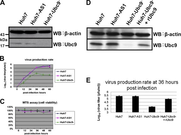
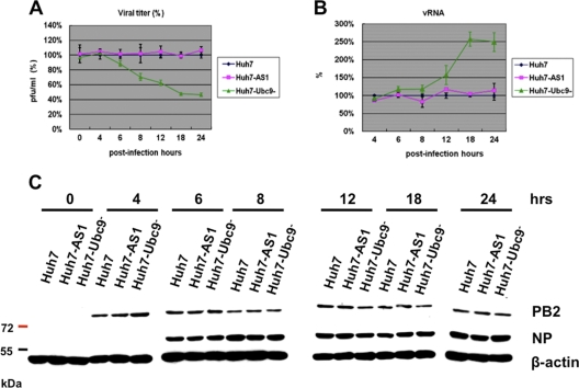
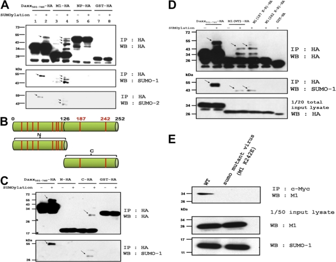
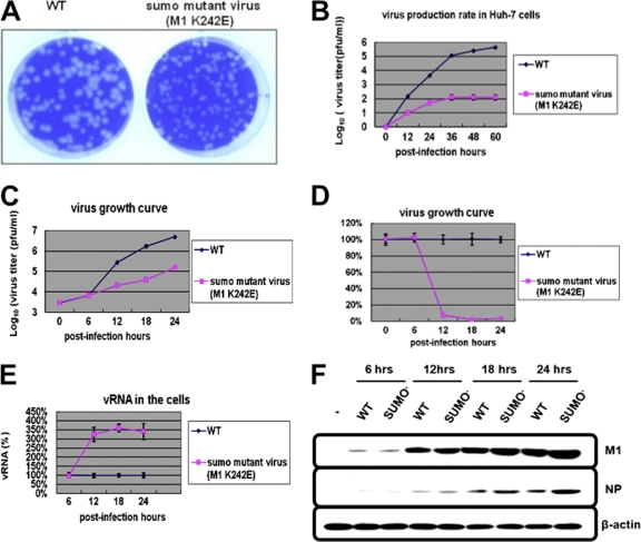
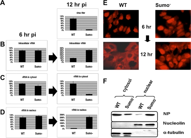
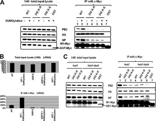
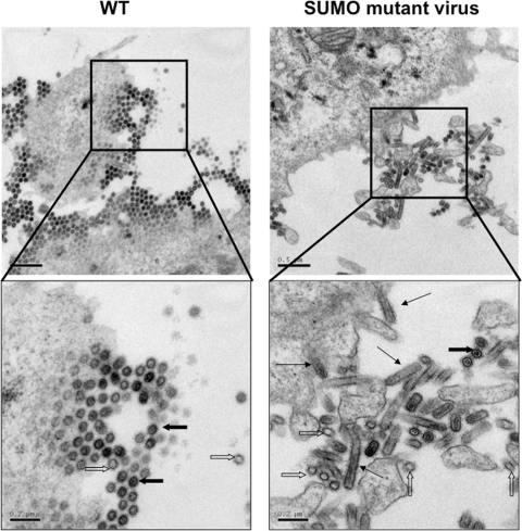
SUMOylation is an important posttranslational modification for regulation of cellular functions and viral replication. Here, we report that protein SUMOylation regulates the replication of influenza A virus at the steps of viral maturation and assembly. Knocking down the SUMO-conjugating enzyme Ubc9 resulted in the reduction of virus production. Dissection of the virus life cycle revealed that SUMOylation is involved in the processes of virus maturation and assembly. The viral matrix protein M1 is SUMOylated at K242. A virus carrying the SUMO-defective M1 produced a lower titer of virus, while its viral proteins and viral RNA (vRNA) accumulated in the cells. Furthermore, the mechanistic studies showed that the SUMOylation of M1 is required for the interaction between M1 and viral RNP (vRNP) to form the M1-vRNP complex. The lack of M1 SUMOylation prevented the nuclear export of vRNP and subsequent viral morphogenesis. Taken together, our findings elucidate that the maturation and assembly of influenza A virus is controlled by the SUMO modification of M1 protein. Therefore, we suggest that M1 can serve as a target for developing a new generation of drugs for flu therapy.


Figures

Fig. 1.
Fig. 1.
Virus production in Ubc9-knocked-down cells. (A) Western blot analysis of Ubc9 in Ubc9-knocked-down and control cells. Cell lysates were prepared from Ubc9-knocked-down cells (Huh7-Ubc9), lentivirus control cells (Huh7-AS1), and naïve Huh7 cells and then subjected to SDS-PAGE. Filter was probed with anti-Ubc9 and anti-actin monoclonal antibodies (MAbs). (B) Comparison of virus production rates. The three cell lines mentioned above were infected with influenza A virus (A/WSN/33) at an MOI of 0.01. Culture fluids were collected at different time points postinfection as indicated, and virus titer was determined by plaque assay on MDCK cells. (C) Cell cytotoxicity assay by MTS assay. (D, E) The rescue experiment for Ubc9 knockdown cells. Huh7-Ubc9 cells were transfected with rUbc9 plasmid, which is a Ubc9 wobble mutant. At 48 h posttransfection, cells were infected with influenza A virus (A/WSN/33) at an MOI of 0.01. At 36 h after infection, total cell lysate was collected and analyzed for protein expression of Ubc9 and actin by Western blot analysis. The virus titer in the culture fluid was determined by plaque assay on MDCK cells. Values represent the means of results from three independent experiments in triplicate, and error bars indicate the standard deviations of the means.
Fig. 2.
Fig. 2.
Silencing of the SUMOylation system affects virus production at the step of virus assembly. The cells as in Fig. 1 were analyzed at different time points postinfection. (A) Analysis of virus production rate. Viral titers were determined by plaque assay on MDCK cells. The wild-type virus titer is normalized as 100%. (B) Quantitative RT-PCR analysis of intracellular vRNA. The primers used for reverse transcription and PCR are described in Material and Methods. Values represent the means of results from two independent experiments in duplicate, and error bars indicate the standard deviations of the means. (C) The expression levels of intracellular viral proteins by immunoblotting.
Fig. 3.
Fig. 3.
In vivo SUMOylation of viral proteins. (A) Plasmids expressing HA-tagged M1 and NP of A/WSN/33 were cotransfected with or without Ubc9- and SUMO-expressing plasmids into HEK293T cells. At 48 h posttransfection, the lysates were immunoprecipitated with anti-HA agarose, followed by Western blot analysis using antibody against HA and SUMO, respectively (20). The arrows indicate the proteins detected by the SUMO antibodies. (B) Schematic representation of conserved lysine residues. Red lines indicate the conserved lysine residues based on Furuse et al. (9). The N and C termini were divided at amino acid 126. (C) In vivo SUMOylation analysis of the truncated M1 proteins. The protocol was the same as that described for panel A. (D) Site-directed mutagenesis analysis of the SUMOylation site on M1. K187R and K242R mutants and the WT of M1 were analyzed as described for panel A. Daxx501-740 and GST serve as positive and negative controls, respectively. The arrows indicate the SUMOylated proteins of Daxx501-740 or M1. (E) HEK293T cells were cotransfected with Ubc9 and c-Myc-tagged SUMO-1 plasmid. At 40 h posttransfection, cells were infected with the WT and sumo mutant viruses at an MOI of 5. At 8 hpi, cell lysates were prepared and subjected to immunoprecipitation with anti-c-Myc agarose and immunoblotted using antibody against M1.
Fig. 4.
Fig. 4.
Characterization of a SUMOylation-defective mutant of A/WSN/33 virus. (A) Plaque morphology of the wild-type and mutant viruses on the monolayer of MDCK cells. (B) Virus production rate of virus-infected Huh7 cells (MOI of 0.01). The virus titer was determined by plaque assay on MDCK cells. (C to F) The various viral parameters in Huh7 cells at various time points after infection with virus at an MOI of 5. (C and D) The virus production rate from infected Huh7 cells. (E) The levels of intracellular vRNA. The RNA was determined by the same way as that described for Fig. 2B. Values represent the means of results from three independent experiments in duplicate, and error bars indicate the standard deviations of the means. (F) The intracellular viral proteins were determined by immunoblotting using antibodies against M1, NP, and β-actin.
Fig. 5.
Fig. 5.
SUMOylated M1 modulates nuclear export of vRNP. Huh7 cells were infected with the WT and sumo mutant viruses at an MOI of 5. (A) Virus production at 12 h postinfection. (B, C, and D) The relative amounts of vRNA in whole-cell, cytosol, and nuclear fractions at 6 and 12 h postinfection. The level of wild-type viral RNA is set at 100%. Values represent the means of results from three independent experiments in duplicate, and error bars indicate the standard deviations of the means. (E) Immunostaining with anti-NP antibody at 6 and 12 h postinfection. (F) The distribution of NP in cytoplasmic and nuclear fractions at 12 h postinfection. Nucleolin and α-tubulin serve as loading controls of nuclear and cytoplasmic fractions, respectively.
Fig. 6.
Fig. 6.
The formation of the M1-vRNP complex. (A) Plasmids expressing various c-Myc-tagged M1 (WT, 242K-R, and 242K-E) were cotransfected with or without Ubc9- and SUMO-expressing plasmids into HEK293T cells. At 40 h posttransfection, cells were infected with WT virus at an MOI of 5. At 8 hpi, cells were harvested and lysates were immunoprecipitated with c-Myc, followed by Western blot analysis using antibody against PB2, HA, NP, and c-Myc. (B) Quantitative RT-PCR analysis of the amount of vRNA coprecipitated with c-Myc-M1. GST-Myc served as a negative control. The amount of vRNA coprecipitated with wild-type M1 is set at 100%. Values represent the means of results from three independent experiments, and error bars indicate the standard deviations of the means. (C) Huh7 and Huh7-Ubc9 cells were transfected with c-Myc-tagged M1 (WT, 242K-R, or 242K-E) plasmids and analyzed as described for panel A. GST-Myc served as a negative control.
Fig. 7.
Fig. 7.
Viral morphology as examined by transmission electron microscopy. Huh7 cells grown on ACLAR embedding film were infected with wild-type or sumo mutant viruses at an MOI of 5. At 12 h postinfection, the monolayer of infected cells was processed for electron microscopy. An 80-nm-thin section was examined. Thick black arrows, normal virus particles; white arrows, empty virus-like particles; thin black arrows, tubular virus particles.

Similar articles

Cited by

References

    1. Ali A., Avalos R. T., Ponimaskin E., Nayak D. P. 2000. Influenza virus assembly: effect of influenza virus glycoproteins on the membrane association of M1 protein. J. Virol. 74:8709–8719 - PMC - PubMed
    1. Arzt S., Petit I., Burmeister W. P., Ruigrok R. W., Baudin F. 2004. Structure of a knockout mutant of influenza virus M1 protein that has altered activities in membrane binding, oligomerisation and binding to NEP (NS2). Virus Res. 99:115–119 - PubMed
    1. Avalos R. T., Yu Z., Nayak D. P. 1997. Association of influenza virus NP and M1 proteins with cellular cytoskeletal elements in influenza virus-infected cells. J. Virol. 71:2947–2958 - PMC - PubMed
    1. Baudin F., Petit I., Weissenhorn W., Ruigrok R. W. 2001. In vitro dissection of the membrane and RNP binding activities of influenza virus M1 protein. Virology 281:102–108 - PubMed
    1. Boggio R., Chiocca S. 2006. Viruses and sumoylation: recent highlights. Curr. Opin. Microbiol. 9:430–436 - PMC - PubMed

Publication types

MeSH terms

LinkOut - more resources