. 2011 Jul;85(13):6669-77.
doi: 10.1128/JVI.00204-11. Epub 2011 Apr 20.

A5-positive primary sensory neurons are nonpermissive for productive infection with herpes simplex virus 1 in vitro

Affiliations

A5-positive primary sensory neurons are nonpermissive for productive infection with herpes simplex virus 1 in vitro

Andrea S Bertke et al. J Virol. 2011 Jul.

Abstract

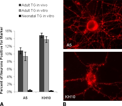
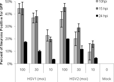
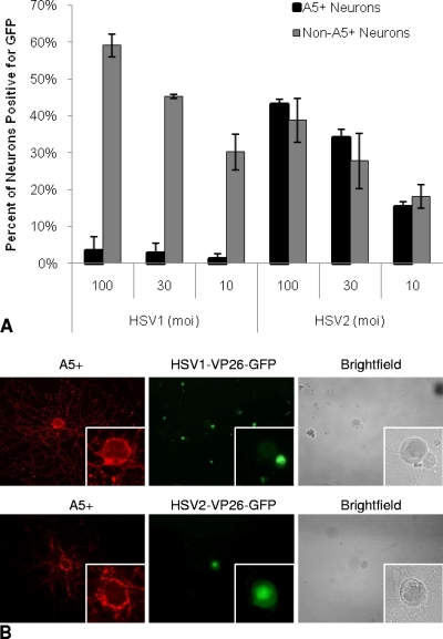
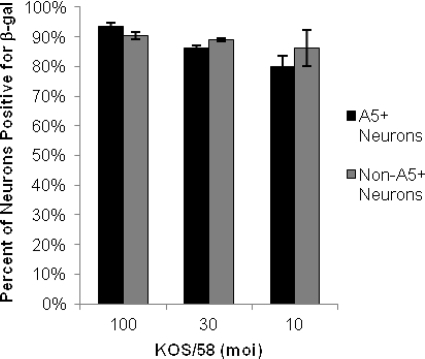
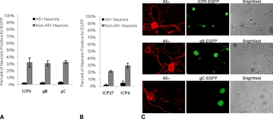
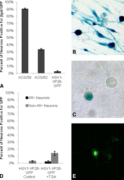
Herpes simplex viruses 1 and 2 (HSV-1 and HSV-2) establish latency and express the latency-associated transcript (LAT) preferentially in different murine sensory neuron populations, with most HSV-1 LAT expression in A5(+) neurons and most HSV-2 LAT expression in KH10(+) neurons. To study the mechanisms regulating the establishment of HSV latency in specific subtypes of neurons, cultured dissociated adult murine trigeminal ganglion (TG) neurons were assessed for relative permissiveness for productive infection. In contrast to that for neonatal TG, the relative distribution of A5(+) and KH10(+) neurons in cultured adult TG was similar to that seen in vivo. Productive infection with HSV was restricted, and only 45% of cultured neurons could be productively infected with either HSV-1 or HSV-2. A5(+) neurons supported productive infection with HSV-2 but were selectively nonpermissive for productive infection with HSV-1, a phenomenon that was not due to restricted viral entry or DNA uncoating, since HSV-1 expressing β-galactosidase under the control of the neurofilament promoter was detected in ∼90% of cultured neurons, with no preference for any neuronal subtype. Infection with HSV-1 reporter viruses expressing enhanced green fluorescent protein (EGFP) from immediate early (IE), early, and late gene promoters indicated that the block to productive infection occurred before IE gene expression. Trichostatin A treatment of quiescently infected neurons induced productive infection preferentially from non-A5(+) neurons, demonstrating that the nonpermissive neuronal subtype is also nonpermissive for reactivation. Thus, HSV-1 is capable of entering the majority of sensory neurons in vitro; productive infection occurs within a subset of these neurons; and this differential distribution of productive infection is determined at or before the expression of the viral IE genes.


Figures

Fig. 1.
Fig. 1.
A5+ and KH10+ neuron distribution in uninfected neuron cultures. (A) Percentages of A5+ and KH10+ adult murine trigeminal neurons in vivo (tissue sections), dissociated adult trigeminal neurons in cultures in vitro, and dissociated neonatal trigeminal neurons in cultures in vitro. (B) Representative fluorescent microscopy images of cultured adult trigeminal neurons positive for the A5 (top) and KH10 (bottom) markers, revealed by use of monoclonal antibodies and a rhodamine-labeled secondary antibody.
Fig. 2.
Fig. 2.
Percentages of neurons expressing GFP following infection with HSV1-VP26-GFP or HSV2-VP26-GFP. Neuronal cultures were infected with HSV1-VP26-GFP or HSV2-VP26-GFP (at an MOI of 100, 30, or 10) for 10, 15, or 24 h and were assayed for GFP expression as a marker of productive infection. Both viral constructs express a VP26-GFP fusion protein during active viral assembly, permitting GFP visualization during productive infection. Results reflect data from six separate experiments for each virus for the 10- and 15-h time points and from two experiments for each virus for the 24-h time point. For each time point, 2,636 to 12,027 neurons were assayed.
Fig. 3.
Fig. 3.
(A) Percentages of A5+ and non-A5+ neurons expressing GFP following infection with HSV1-VP26-GFP or HSV2-VP26-GFP. Neuronal cultures were infected with HSV1-VP26-GFP or HSV2-VP26-GFP. A5+ and non-A5+ neurons were evaluated for GFP expression as a marker of productive infection at 10 h postinfection. Results reflect data from two separate experiments for each MOI. A minimum of 3,520 total neurons were assayed for each MOI. (B) Fluorescent microscope images of A5+ neurons (red) with HSV1-VP26-GFP (top) or HSV2-VP26-GFP (bottom) productive infection (green). (Insets) Higher-magnification images of A5+ neurons. HSV1-VP26-GFP productive infection, indicated by GFP expression, was frequently found in non-A5+ neurons adjacent to nonproductively infected A5+ neurons, while HSV2-VP26-GFP was found in A5+ neurons as well as in non-A5+ neurons.
Fig. 4.
Fig. 4.
Percentage of neurons expressing β-galactosidase following infection with KOS/58. Expression of β-gal, driven by the neurofilament promoter at the HSV-1 gC locus, was detected in A5+ and non-A5+ neurons by immunofluorescence with MAb A5 and an anti-β-gal polyclonal antibody. Experiments were carried out in duplicate, and a minimum of 3,726 neurons were assayed for each MOI.
Fig. 5.
Fig. 5.
Viral gene expression in A5+ and non-A5+ neurons. Neuronal cultures were infected at an MOI of 30 with viral constructs expressing EGFP under the control of immediate early (ICP0), early (gB), and late (gC) gene promoters, inserted at the gC locus. A5+ and non-A5+ neurons were evaluated for EGFP expression. (A) EGFP expression was determined for RE-pICP0-EGFP at 6 h, for RE-pgB-EGFP at 8 h, and for RE-pgC-EGFP at 10 h. Results reflect data from five separate experiments with ∼4,100 to 9,300 neurons assayed for each virus studied. (B) EGFP expression was determined for RE-pICP27-EGFP and RE-pICP4-EGFP at 6 h. Results reflect data from three separate experiments with ∼4,100 to 4,200 neurons assayed for each virus studied. (C) Fluorescent microscope images of A5+ neurons (red) with EGFP expression (green) after infection with RE-pICP0-EGFP (top), RE-pgB-EGFP (center), or RE-pgC-EGFP (bottom). Corresponding bright-field images are shown on the right.
Fig. 6.
Fig. 6.
Viral gene expression from KOS/58, KOS/62, and HSV1-VP26-GFP. (A) Neuronal cultures were infected with either KOS/58, KOS/62, or HSV1-VP26-GFP (at an MOI of 10) and were evaluated for β-gal or GFP expression 5 days postinoculation. (B) β-gal-positive neurons (blue) 5 days after infection with KOS/58 (β-gal from the neurofilament light promoter). (C) β-gal-positive neurons (blue) 5 days after infection with KOS/62 (β-gal from the LAT promoter). (D) Five days after infection with HSV1-VP26-GFP (at an MOI of 10), cultures were treated with TSA or with a control medium containing no TSA; 24 h later, A5+ and non-A5+ neurons were evaluated for GFP expression. (E) GFP expression in a cultured neuron 24 h after TSA treatment of a culture quiescently infected with HSV1-VP26-GFP. Results reflect data collected from two or more separate experiments with a minimum of 1,503 total neurons assayed for each virus.

References

    1. Arthur J. L., et al. 2001. Herpes simplex virus type 1 promoter activity during latency establishment, maintenance, and reactivation in primary dorsal root neurons in vitro. J. Virol. 75:3885–3895 - PMC - PubMed
    1. Bedadala G. R., Pinnoji R. C., Hsia S. C. 2007. Early growth response gene 1 (Egr-1) regulates HSV-1 ICP4 and ICP22 gene expression. Cell Res. 17:546–555 - PMC - PubMed
    1. Bennett D. L., Averill S., Clary D. O., Priestley J. V., McMahon S. B. 1996. Postnatal changes in the expression of the trkA high-affinity NGF receptor in primary sensory neurons. Eur. J. Neurosci. 8:2204–2208 - PubMed
    1. Bennett D. L., et al. 1998. A distinct subgroup of small DRG cells express GDNF receptor components and GDNF is protective for these neurons after nerve injury. J. Neurosci. 18:3059–3072 - PMC - PubMed
    1. Bertke A. S., et al. 2009. Latency-associated transcript (LAT) exon 1 controls herpes simplex virus species-specific phenotypes: reactivation in the guinea pig genital model and neuron subtype-specific latent expression of LAT. J. Virol. 83:10007–10015 - PMC - PubMed

Publication types

MeSH terms