. 2011 Jul;85(13):6343-52.
doi: 10.1128/JVI.00101-11. Epub 2011 Apr 20.

RNA elements directing translation of the duck hepatitis B Virus polymerase via ribosomal shunting

Affiliations

RNA elements directing translation of the duck hepatitis B Virus polymerase via ribosomal shunting

Feng Cao et al. J Virol. 2011 Jul.

Abstract

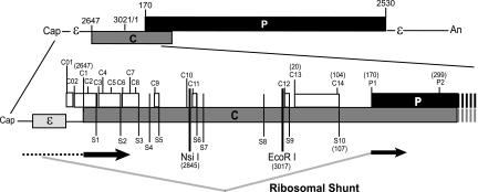
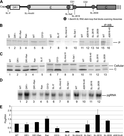
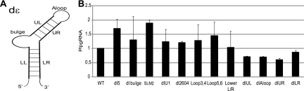
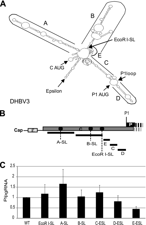
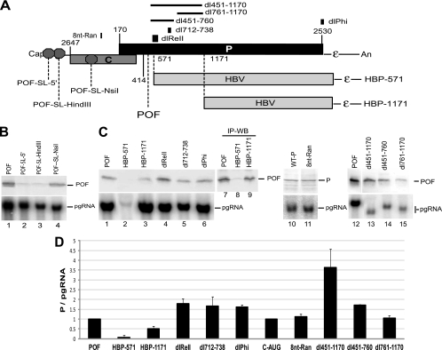
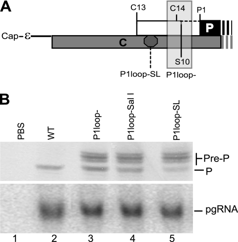
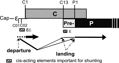
The duck hepatitis B virus (DHBV) reverse transcriptase (P) is translated from the downstream position on a bicistronic mRNA, called the pregenomic RNA, through a poorly characterized ribosomal shunt. Here, the positions of the discontinuous ribosomal transfer during shunting were mapped, and RNA elements important for shunting were identified as a prelude to dissecting the shunting mechanism. Mutations were introduced into the DHBV genome, genomic expression vectors were transfected into cells which support reverse transcription, and P translation efficiency was defined as the ratio of P/mRNA. Five observations were made. First, ribosomes departed from sequences that comprise the RNA stem-loop called ε that is key to viral replication, but the known elements of ε were not needed for shunting. Second, at least two landing sites for ribosomes were found on the mRNA. Third, all sequences upstream of ε, most sequences between the cap and the P AUG, and sequences within the P-coding region were dispensable for shunting. Fourth, elements on the mRNA involved in reverse transcription or predicted to be involved in shunting on the basis of mechanisms documented in other viruses, including short open reading frames near the departure site, were not essential for shunting. Finally, two RNA elements in the 5' portion of the mRNA were found to assist shunting. These observations are most consistent with shunting being directed by signals that act through an uncharacterized RNA secondary structure. Together, these data indicate that DHBV employs either a novel shunting mechanism or a major variation on one of the characterized mechanisms.


Figures

Fig. 1.
Fig. 1.
Organization of the DHBV3 pgRNA. The pgRNA is 3.3-kb capped and polyadenylated mRNA with a terminal redundancy of approximately 270 nt. The top section shows the relative positions of the major ORFs and functional elements on the pgRNA. The bottom section shows an expanded view of the 5′ end of the RNA, with the location of the features relevant to this study displayed. The genomic positions of the elements are indicated for DHBV strain 3 (nucleotides 3021 and 1 are adjacent to each other within the unique EcoRI site on the circular DNA form of the genome). The ribosomal shunt as it was known at the start of this study is shown at the bottom, with regions that are scanned by ribosomes shown with a dashed line, regions that are translated shown as thick black lines, and the ribosomal shunt shown with gray lines.
Fig. 2.
Fig. 2.
Epsilon sequences contain the donor site, and an element important for shunting is between ε and the C1 AUG. LMH cells were transfected with mutant DHBV genomic expression constructs, and pgRNA and P levels were detected by Northern and Western blotting (WB) of lysates on day 1 posttransfection. P translation efficiency was defined as the amount of P divided by the amount of pgRNA in the same lysate. (A) Positions of relevant elements within the 5′ end of the pgRNA. Octagons represent the positions at which the BamHI-SL was inserted to block scanning ribosomes. (B) Representative Western blots for P. (C) Representative Western blots for C. (D) Representative Northern blots for the pgRNA. (E) P translation efficiency for the mutants with lesions in the 5′ end of the pgRNA. Data are normalized to the activity of the wild-type (WT) construct and shown as the mean ± standard deviation from three to five replicate experiments. IP, immunoprecipitation.
Fig. 3.
Fig. 3.
Proper folding of ε and the known functional elements of ε are not needed for shunting. LMH cells were transfected with DHBV genomic expression constructs carrying mutations in ε, and pgRNA and P levels were detected by Northern and Western blotting of lysates on day 1 posttransfection. (A) The structure of DHBV ε showing regions that were deleted or mutated: apical loop (Aloop), upper left stem (UL), Bulge, lower left stem (LL), upper right stem (UR), and lower right stem (LR). (B) P translation efficiency from the mutant constructs shown as the mean ± standard deviation of three to five independent experiments.
Fig. 4.
Fig. 4.
Only element E between the C1 and P1 AUGs contributes to shunting. (A) A predicted fold for the 5′ end of the DHBV pgRNA. This predicted fold shows the genomic regions defined as A through E; it was used to guide a scanning deletion analysis of the sequences upstream of the P1 AUG. (B) Relative positions of the deletions and the insertions of the BamHI-SL within the 5′ UTR. (C) P translation efficiency from DHBV genomic expression constructs carrying deletions of the A to E elements. Elements A to E were sequentially deleted. The BamHI-SL was inserted into the A and B deletion sites and into the EcoRI site in the C, D, and E deletion mutants to block fortuitous activation of ribosomal scanning. The mutants were transfected into LMH cells, and P translation efficiency was calculated. The results are shown as the mean ± standard deviations from three independent experiments.
Fig. 5.
Fig. 5.
Sequences downstream of the P1 AUG are not needed for shunting. Mutations were introduced into genomic expression vectors in the D1.5G-POF or D1.5G background, the mutants were transfected into LMH cells, and POF, P, and pgRNA levels were measured by Western and Northern analysis of the same lysates 1 day posttransfection. (A) Structure of the pgRNA showing the locations of the elements altered in this experiment. The stop codon defining the POF truncated form of P is shown, and octagons represent the insertion sites for the BamHI-SL used to block scanning ribosomes. The regions replaced with HBV sequences are shown. (B) Northern and Western blots showing that the POF fragment is translated by ribosomal shunting. (C) Representative Western and Northern blots. (D) P translation efficiency for the mutants expressed as the mean ± standard deviation from three to five independent experiments.
Fig. 6.
Fig. 6.
Ribosomes land at more than one acceptor site. Mutations were introduced into genomic expression vectors in the P1loop− background to permit synthesis of the pre-P protein from DHBV strain 3, the mutants were transfected into LMH cells, and P, pre-P, and pgRNA levels were measured by Western and Northern analysis of the same lysates 1 day posttransfection. (A) Structure of the pgRNA showing the locations of the elements altered in this experiment. (B) Representative Western blot (upper panel) and Northern blot (lower panel) showing accumulation of P, pre-P, and the pgRNA. PBS, pBluescript vector.
Fig. 7.
Fig. 7.
Summary of the DHBV P shunt and the RNA elements directing ribosomal transfer.

Similar articles

Cited by

References

    1. Calvert J., Summers J. 1994. Two regions of an avian hepadnavirus RNA pregenome are required in cis for encapsidation. J. Virol. 68:2084–2090 - PMC - PubMed
    1. Cao F., Scougall C. A., Jilbert A. R., Tavis J. E. 2009. Pre-P is a secreted glycoprotein encoded as an N-terminal extension of the duck hepatitis B virus polymerase gene. J. Virol. 83:1368–1378 - PMC - PubMed
    1. Chang C., Hirsch R. C., Ganem D. 1995. Sequences in the preC region of duck hepatitis B virus affect pregenomic RNA accumulation. Virology 207:549–554 - PubMed
    1. Condreay L. D., Aldrich C. E., Coates L., Mason W. S., Wu T. T. 1990. Efficient duck hepatitis B virus production by an avian liver tumor cell line. J. Virol. 64:3249–3258 - PMC - PubMed
    1. de Breyne S., Simonet V., Pelet T., Curran J. 2003. Identification of a cis-acting element required for shunt-mediated translational initiation of the Sendai virus Y proteins. Nucleic Acids Res. 31:608–618 - PMC - PubMed

Publication types

MeSH terms

LinkOut - more resources