. 2011 Jul;85(13):6185-96.
doi: 10.1128/JVI.00040-11. Epub 2011 Apr 20.

RNA splicing in a new rhabdovirus from Culex mosquitoes

Affiliations

RNA splicing in a new rhabdovirus from Culex mosquitoes

Ryusei Kuwata et al. J Virol. 2011 Jul.

Abstract

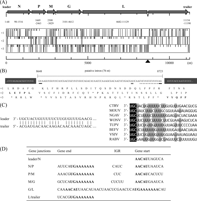
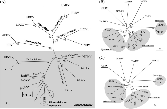
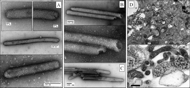
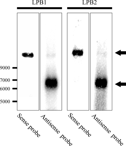
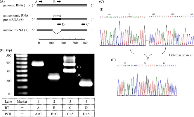
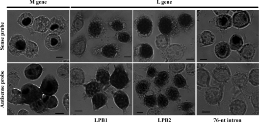
Among members of the order Mononegavirales, RNA splicing events have been found only in the family Bornaviridae. Here, we report that a new rhabdovirus isolated from the mosquito Culex tritaeniorhynchus replicates in the nuclei of infected cells and requires RNA splicing for viral mRNA maturation. The virus, designated Culex tritaeniorhynchus rhabdovirus (CTRV), shares a similar genome organization with other rhabdoviruses, except for the presence of a putative intron in the coding region for the L protein. Molecular phylogenetic studies indicated that CTRV belongs to the family Rhabdoviridae, but it is yet to be assigned a genus. Electron microscopic analysis revealed that the CTRV virion is extremely elongated, unlike virions of rhabdoviruses, which are generally bullet shaped. Northern hybridization confirmed that a large transcript (approximately 6,500 nucleotides [nt]) from the CTRV L gene was present in the infected cells. Strand-specific reverse transcription-PCR (RT-PCR) analyses identified the intron-exon boundaries and the 76-nt intron sequence, which contains the typical motif for eukaryotic spliceosomal intron-splice donor/acceptor sites (GU-AG), a predicted branch point, and a polypyrimidine tract. In situ hybridization exhibited that viral RNAs are primarily localized in the nucleus of infected cells, indicating that CTRV replicates in the nucleus and is allowed to utilize the host's nuclear splicing machinery. This is the first report of RNA splicing among the members of the family Rhabdoviridae.


Figures

Fig. 1.
Fig. 1.
Genome organization of CTRV. (A) The upper diagram represents the schematic transcription map of the 11,190-nt CTRV positive-strand antigenome. The shaded arrows indicate the positions of the ORFs for the five typical rhabdovirus proteins, N, P, M, G, and L. The numbers below the arrows indicate the sequence positions of the leader, ORFs, and trailer. The lower diagram represents the positions of start codons (short vertical lines) and stop codons (long vertical lines) in each of the three reading frames on the positive strand. Note that the coding region for the CTRV L protein is interrupted by an in-frame stop codon around sequence position 8700 (black arrowhead). (B) Antigenomic nucleotide sequences (upper) and predicted amino acid sequences of the three reading frames (lower) around the putative intron in the L gene. The 76-nt intron (8648 to 8723 nt) contains typical splice site motifs—GU-AG boundaries (underlines), a predicted branch point (double underline), and a polypyrimidine tract (dotted line). If the intron is spliced out, the translational reading frame shifts from +2 to +3 to generate a continuous L protein. (C) Complementarity of the 3′ and 5′ termini of the CTRV genome (left) and an alignment of the leader sequence of CTRV with those of other animal-infecting rhabdoviruses (right). The conserved 3′ terminal trinucleotides UGC are indicated in reverse type. The nucleotides shaded in gray indicate more than 50% sequence identity. (D) Sequences of the gene junctions in the CTRV antigenomic RNA. The upstream gene end sequences, IGRs, and the downstream gene start sequences are indicated. Consensus sequences of the predicted transcription start/stop signals are shown in bold. Note that the transcription start signal of the L gene precedes the transcription stop signal of G gene, resulting in a 36-nt gene overlap at the G/L junction.
Fig. 2.
Fig. 2.
Phylogenetic relationships between CTRV and other NNS RNA viruses. These dendrograms were constructed based on the protein sequence similarities of the highly conserved domain III of the L protein among selected members of the order Mononegavirales (A) and the family Rhabdoviridae (B) or based on the protein sequence similarities of partial N protein among selected members of the family Rhabdoviridae (C). In dendrogram A, family or subfamily names (bold face) and genus names are indicated on each appropriate branch. In dendrograms B and C, the genus names in the family Rhabdoviridae are underlined, and the viruses in the same genus are shaded. More than 50% of bootstrap values from 1,000 intermediate trees are indicated at the branch points. The bar represents the expected substitutions per site. Abbreviations of NNS RNA virus and sequence accession numbers (L and/or N protein) used in this study are follows: ABDV, avian Borna disease virus (ACS32310); ARV, Adelaide river virus (AF234534 and U10363); BDV, Borna disease virus (NC_001607); BEFV, bovine ephemeral fever virus (AF234533); COCV, Cocal virus (ACB47438 and ACB47434); DAffSV, Drosophila affinis sigma virus (ACU65445); DMelSV (SIGMAV), Drosophila melanogaster sigma virus (NC_013135); DObsSV, Drosophila obscura sigma virus (ACU65444 and ACU65439); EBOV, Zaire ebolavirus (NC_002549); FLAV, Flanders virus (AH012179); HMPV, human metapneumovirus (NC_004148); HPIV1, human parainfluenza virus 1 (NC_003461); HRSV, human respiratory syncytial virus (NC_001781); IHNV, infectious hematopoietic necrosis virus (NC_001652); ISFV, Isfahan virus (AJ810084); LNYV, lettuce necrotic yellows virus (NC_007642); MARV, Lake Victoria marburgvirus (NC_001608); MOKV, Mokola virus (NC_006429); MOUV, Moussa virus (FJ985748), NCMV, northern cereal mosaic virus (AB030277); NDV, Newcastle disease virus (NC_002617); NGAV, Ngaingan virus (NC_013955); PEFV, pike fry rhabdovirus (FJ872827); RABV, rabies virus (GU345748); RYSV, rice yellow stunt virus (AB011257); SCRV, Siniperca chuatsi rhabdovirus (NC_008514); SFRV, starry flounder rhabdovirus (AY450644); SVCV, spring viremia of carp virus (NC_002803); SYNV, sonchus yellow net virus, (NC_001615); TUPV, tupaia rhabdovirus (AY840978); VHSV, viral hemorrhagic septicemia virus (NC_000855); VSIV, vesicular stomatitis Indiana virus (NC_001560); WONV, Wongabel virus (NC_011639).
Fig. 3.
Fig. 3.
Electron microscopic analyses of CTRV virions and thin-section of infected C6/36 cells. (A) The virions exhibited width uniformity and substantial variation in length, which are straight with two rounded tips and surrounded by a layer with projections entirely on the virion surface. The penetrating stain image showed the periodic fringe of the internal structure. (B) The truncated virion image shows the presence of a hollow cavity in the interior of the particle. (C) Some parts of the virion may be seen without the outer envelope, which seems to expose the naked inner structure. (D) Thin section of the C6/36 cells infected with CTRV. Abbreviations: N, nucleus; MT; mitochondrion. White arrows indicate the CTRV virions in the intracellular vacuoles, and the lower panel shows detail in dotted-line box in the upper panel. Scale bar, 100 nm.
Fig. 4.
Fig. 4.
Northern hybridization analysis of the CTRV L gene transcript in infected cells. Total RNA from CTRV-infected C6/36 cells was electrophoresed in a 0.8% denaturing formaldehyde agarose gel. Hybridization was performed using sense or antisense RNA probes specific for the regions upstream (LBP1; sequence position 4894 to 5752) or downstream (LBP2; sequence position 10172 to 11125) of the putative intron in the L gene. Black arrows indicate the hybridizing bands corresponding to the expected sizes of viral genomic or antigenomic RNA (upper) and viral full-length L mRNA (lower), respectively. Both a single-stranded RNA ladder (New England BioLabs, Ipswich, MA) and an RNA 6000 Ladder (Ambion Inc., Austin, TX) were used as molecular weight markers.
Fig. 5.
Fig. 5.
Strand-specific RT-PCR analysis for the CTRV L gene transcript. (A) Schematic map of partial regions of CTRV genomic RNA (negative strand; −), antigenomic RNA, and mRNA (positive strand; +) around the intron in the L gene, as shown with a scale bar. The four black arrows indicate the positions and orientations of CTRV gene-specific primers used for strand-specific RT-PCR. For RT, primers A and B anneal specifically to negative-strand RNA, whereas primers C and D anneal specifically to positive-strand RNA, respectively. (B) Agarose gel electrophoresis of the strand-specific RT-PCR products (lanes 1 to 4). The lower table indicates the primers used for each RT and the following PCR. Note that the smallest fragment (II) on lane 3 is an amplified product from spliced mature mRNA with the expected size of 297 bp. (C) Sequence chromatograms of the two PCR products (fragments I and II) in panel B, which were directly sequenced with primer A. The upper chromatogram was obtained from fragment I, and the lower one was obtained from fragment II. Note that the sequence of fragment II contains a deletion of 76 nt, as expected in Fig. 1B.
Fig. 6.
Fig. 6.
Subcellular localization of CTRV RNA in the C6/36 cells persistently infected with CTRV. DIG-labeled sense or antisense RNA probe for CTRV M and L genes specifically recognized negative-sense or positive-sense viral RNA, respectively. Several uninfected cells were also seen in the visual field. Scale bar, 5 μm.

Similar articles

Cited by

References

    1. Ammar el-D., Tsai C. W., Whitfield A. E., Redinbaugh M. G., Hogenhout S. A. 2009. Cellular and molecular aspects of rhabdovirus interactions with insect and plant hosts. Annu. Rev. Entomol. 54:447–468 - PubMed
    1. Bock J. O., Lundsgaard T., Pedersen P. A., Christensen L. S. 2004. Identification and partial characterization of Taastrup virus: a newly identified member species of the Mononegavirales. Virology 319:49–59 - PubMed
    1. Boulo S., Akarsu H., Ruigrok R. W., Baudin F. 2007. Nuclear traffic of influenza virus proteins and ribonucleoprotein complexes. Virus Res. 124:12–21 - PubMed
    1. Bourhy H., Cowley J. A., Larrous F., Holmes E. C., Walker P. J. 2005. Phylogenetic relationships among rhabdoviruses inferred using the L polymerase gene. J. Gen. Virol. 86:2849–2858 - PubMed
    1. Chen B. J., Lamb R. A. 2008. Mechanisms for enveloped virus budding: can some viruses do without an ESCRT? Virology 372:221–232 - PMC - PubMed

Publication types

MeSH terms