. 2011 Jul;85(13):6148-61.
doi: 10.1128/JVI.02608-10. Epub 2011 Apr 20.

The single RBP-Jkappa site within the LANA promoter is crucial for establishing Kaposi's sarcoma-associated herpesvirus latency during primary infection

Affiliations

The single RBP-Jkappa site within the LANA promoter is crucial for establishing Kaposi's sarcoma-associated herpesvirus latency during primary infection

Jie Lu et al. J Virol. 2011 Jul.

Abstract

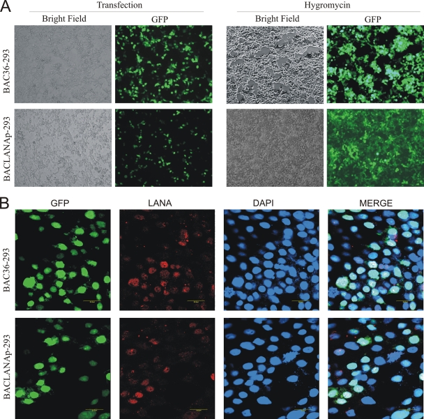
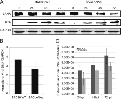
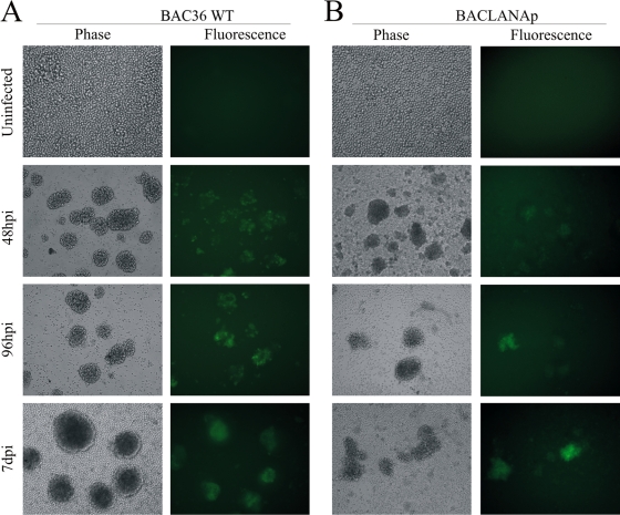
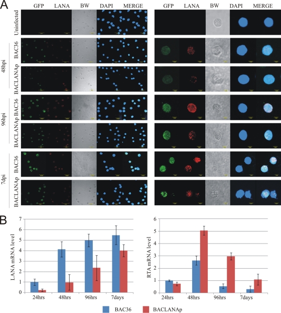
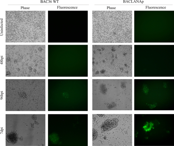
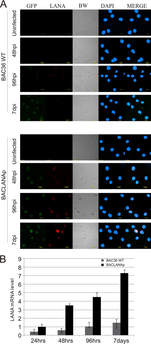
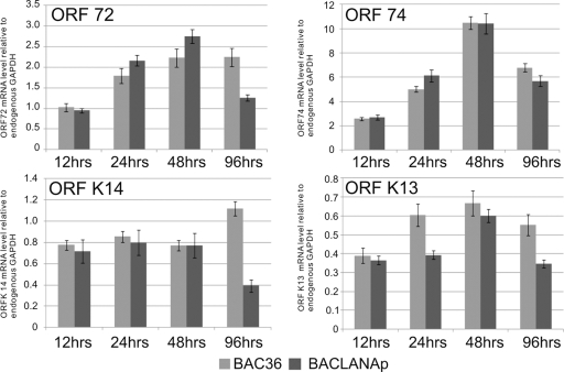
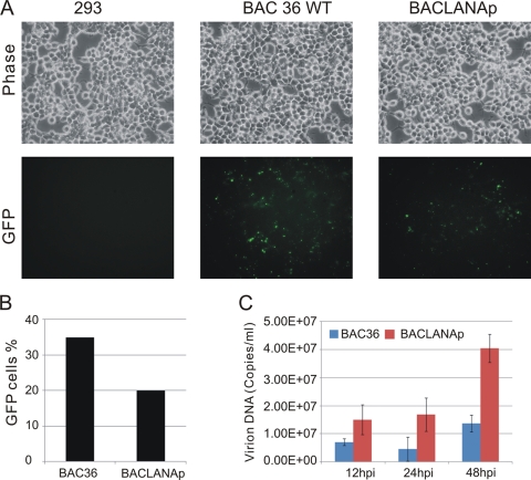
Kaposi's sarcoma-associated herpesvirus (KSHV; or human herpesvirus 8 [HHV8]) is implicated in the pathogenesis of many human malignancies including Kaposi's sarcoma (KS), multicentric Castleman's disease (MCD), and primary effusion lymphoma (PEL). KSHV infection displays two alternative life cycles, referred to as the latent and lytic or productive cycle. Previously, we have reported that the replication and transcription activator (RTA), a major lytic cycle transactivator, contributes to the development of KSHV latency by inducing latency-associated nuclear antigen (LANA) expression during early stages of infection by targeting RBP-Jκ, the master regulator of the Notch signaling pathway. Here, we generated a bacterial artificial chromosome (BAC) KSHV recombinant virus with a deletion of the RBP-Jκ site within the LANA promoter to evaluate the function of the RBP-Jκ cognate site in establishing primary latent infection. The results showed that genetic disruption of the RBP-Jκ binding site within the KSHV LANA promoter led to enhanced expression of the KSHV-encoded immediate early RTA, resulting in an increase in lytic replication during primary infection of human peripheral blood mononuclear cells (PBMCs). This system provides a powerful tool for use in indentifying additional cellular and viral molecules involved in LANA-mediated latency maintenance during the early stages of KSHV infection.


Figures

Fig. 1.
Fig. 1.
Generation of a recombinant KSHV BACmid with the RBP-Jκ binding site deleted within the LANA promoter. (A) Schematic diagram showing generation of a recombinant KSHV BACmid with RBP-Jκ binding site deleted within the LANA promoter, BACLANAp. (B) Agarose gel and Southern blots with BAC36 WT (lane 1) and the mutated BACmid, BACLANAp, with neo cassette (Unfloxed; lane 2) and without neo cassette (Floxed; lane 3), cleaved with XhoI. (C) PCR for BAC36 WT and BACLANAp at the junction. DNA fragments were sequenced, and the mutations were confirmed. EtBr, ethidium bromide.
Fig. 2.
Fig. 2.
Establishment of BAC36 WT and BACLANAp stable cell lines. (A) Cells were transfected by the CaPO4 method with BAC36 WT and BACLANAp. GFP signal was monitored 2 days after transfection; subsequently the cells were passaged and selected with hygromycin B. The homogenous population of GFP-positive cells harboring KSHV BAC36 WT DNA was obtained after 3 to 4 weeks of selection. (B) Immunofluorescence analysis for LANA in BAC36 WT-293 and BACLANAp-293 cells.
Fig. 3.
Fig. 3.
Effect of RBP-Jκ site deletion on the LANA promoter in KSHV lytic life cycle. (A) Comparison of the level of viral gene expression in BAC36 WT-293 and BACLANAp-293 cells. Western blotting for RTA and LANA at 24, 48, and 72 h after lytic induction of BAC36 WT and BACLANAp 293 cells in the presence of butyrate (3 mM) and TPA (20 ng/ml). GAPDH was used as a control to ensure equal loading of each sample. (B) Detection of KSHV viruses using TR primers in the BAC36 WT-293 and BACLANAp-293 cells. Total intracellular DNAs were extracted from cells, and viral genome DNAs were measured by a real-time PCR with the primers to TR from a 100-ng DNA sample with GAPDH as a control. (C) Detection of KSHV viruses using TR primers in the supernatant of BAC36 WT-293 and BACLANAp-293 cells induced by butyrate and TPA. At different time points postinduction (0, 24, 48, and 72 h), extracellular KSHV virions in the supernatant were harvested and concentrated. Virus stocks were treated with DNase for 1 h at 37°C, and the reaction was stopped by addition of a final concentration of 8 mM EDTA and incubation at 75°C for 10 min. Viral DNAs were extracted and analyzed by RT-PCR using primers to TR. hpi, hours postinfection.
Fig. 4.
Fig. 4.
Relative infectivity of KSHV BAC36 WT and BACLANAp viruses. PBMCs were infected by KSHV BAC36 WT and BACLANAp using equal amounts of viruses as determined by copy number. GFP signal was monitored at 48 h, 96 h, and 7 days postinfection.
Fig. 5.
Fig. 5.
Effect of KSHV-infected PBMCs at 48 h, 96 h, and 7 days. (A) Immunofluorescence assay for PBMCs infected by BAC36 WT and BACLANAp viruses. Uninfected and infected PBMCs at 48 h, 96 h, and 7 days were stained for LANA protein expression. (B) Quantitative analysis for determination of KSHV infection in PBMCs. Total RNAs were prepared and transcribed to cDNA after 24 h, 48 h, 96 h, and 7 days postinfection. The qRT-PCR analysis with the primers for LANA and RTA was performed using the Power SYBR green PCR Master Mix with GAPDH as a control. Error bars indicate standard deviations from three separate experiments. BW, phase.
Fig. 6.
Fig. 6.
BACLANAp produces KSHV virus progeny during early infection. PBMCs were infected with BAC36 WT and BACLANAp at specific times postinfection; the supernatant was collected and used to infect new PBMCs. Phase-contrast and fluorescence images of PBMCs reinfected by BAC36 WT and BACLANAp viruses after 48 h, 96 h, and 7 days are shown.
Fig. 7.
Fig. 7.
(A) Immunofluorescence analysis for PBMCs reinfected by BAC36 WT and BACLANAp viruses collected from the supernatant of infected PBMCs at 7 days postinfection. Uninfected and reinfected PBMCs at 48 h, 96 h, and 7 days postinfection were stained for LANA protein expression. (B) Quantitative analysis for determination of KSHV reinfected PBMCs. Total RNAs were prepared and transcribed to cDNA after 24 h, 48 h, 96 h, and 7 days postinfection. qRT-PCR analysis with the primers for LANA was performed using the Power SYBR green PCR Master Mix with GAPDH as a control. Error bars indicate standard deviations from three separate experiments.
Fig. 8.
Fig. 8.
Quantitative analysis of ORFs 72, 74, K13, and K14 expression in KSHV-infected PBMCs at 24 h, 48 h, 96 h, and 7 days. Total RNAs were prepared and transcribed to cDNA after 24 h, 48 h, 96 h, and 7 days postinfection. The qRT-PCR analysis with the primers for ORFs 72, 74, K13, and K14 was performed using the Power SYBR green PCR Master Mix with GAPDH as a control. Error bars indicate standard deviations from two separate experiments.
Fig. 9.
Fig. 9.
Infectivity of BACLANAp recombinant viruses in HEK 293 cells. (A) HEK 293 cells were infected by BACLANAp recombinant viruses. GFP expression was examined under a fluorescence microscope at 48 h postinfection. (B) Counts of 293 cells infected by BACLANAp recombinant viruses (GFP-positive cells). (C) The supernatant was then collected at 12, 24, and 48 h postinfection, viral particles were concentrated, and viral genomic DNAs were quantified by real-time PCR with primers specific for KSHV TR. Error bars indicate standard deviations from two separate experiments.

Similar articles

Cited by

References

    1. An F. Q., et al. 2005. The latency-associated nuclear antigen of Kaposi's sarcoma-associated herpesvirus modulates cellular gene expression and protects lymphoid cells from p16 INK4A-induced cell cycle arrest. J. Biol. Chem. 280:3862–3874 - PubMed
    1. Barcy S., et al. 2008. Gamma delta+ T cells involvement in viral immune control of chronic human herpesvirus 8 infection. J. Immunol. 180:3417–3425 - PubMed
    1. Boivin G., Gaudreau A., Routy J. P. 2000. Evaluation of the human herpesvirus 8 DNA load in blood and Kaposi's sarcoma skin lesions from AIDS patients on highly active antiretroviral therapy. AIDS 14:1907–1910 - PubMed
    1. Borggrefe T., Oswald F. 2009. The Notch signaling pathway: transcriptional regulation at Notch target genes. Cell. Mol. Life Sci. 66:1631–1646 - PMC - PubMed
    1. Brown E. E., et al. 2006. Virologic, hematologic, and immunologic risk factors for classic Kaposi sarcoma. Cancer 107:2282–2290 - PubMed

Publication types

MeSH terms

Substances

LinkOut - more resources