. 2011 Jul;85(13):6502-12.
doi: 10.1128/JVI.02560-10. Epub 2011 Apr 20.

NF-kappaB-mediated modulation of inducible nitric oxide synthase activity controls induction of the Epstein-Barr virus productive cycle by transforming growth factor beta 1

Affiliations

NF-kappaB-mediated modulation of inducible nitric oxide synthase activity controls induction of the Epstein-Barr virus productive cycle by transforming growth factor beta 1

Lassad Oussaief et al. J Virol. 2011 Jul.

Abstract

Transforming growth factor beta 1 (TGF-β1) signal transduction has been implicated in many second-messenger pathways, including the NF-κB pathway. We provide evidence of a novel TGF-β1-mediated pathway that leads to extracellular signal-regulated kinase (ERK) 1/2 phosphorylation, which in turn induces expression of an Epstein-Barr virus (EBV) protein, ZEBRA, that is responsible for the induction of the viral lytic cycle. This pathway includes two unexpected steps, both of which are required to control ERK 1/2 phosphorylation: first, a quick and transient activation of NF-κB, and second, downregulation of inducible nitric oxide synthase (iNOS) activity that requires the participation of NF-κB activity. Although necessary, NF-κB alone is not sufficient to produce downregulation of iNOS, suggesting that another uncharacterized event(s) is involved in this pathway. Dissection of the steps involved in the switch from the EBV latent cycle to the lytic cycle will be important to understand how virus-host relationships modulate the innate immune system.


Figures

Fig. 1.
Fig. 1.
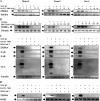
TGF-β1 induces rapid and transient activation of NF-κB in BL cell lines. (A) Mutu-I, Kem-I, and Sav-I cells were pretreated or not with MG262 prior to stimulation with TGF-β1 (2 ng/ml) for 10, 15, or 30 min. Cells were harvested, washed, and lysed, and then equal amounts of protein were separated by SDS-PAGE and analyzed by Western blotting with antibodies to IκBα and tubulin. (B and C) Mutu-I, Kem-I, and Sav-I cells were treated with TGF-β1 (2 ng/ml) for various periods of time. At the indicated time points, cells were harvested; the nuclear (B) and cytoplasmic (C) extracts were isolated, and the content of NF-κB p65 protein was determined by ELISA (Imgenex). Loading of nuclear and cytosolic fractions was assayed by blotting with antibodies to HP1γ (for the nuclear fraction) and tubulin (for the cytosolic fraction).
Fig. 2.
Fig. 2.
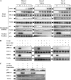
Inhibitors of the NF-κB signaling pathway abolish TGF-β1-induced EBV reactivation. (A and B) Mutu-I, Kem-I, and Sav-I cells were treated with increasing concentrations of BAY11-7082 (0.5, 1, 2, 5, or 10 μM) (A) or IKK inhibitor V (10, 20, 50, or 100 nM) (B) for 1 h prior to incubation with TGF-β1 (2 ng/ml). Seventeen hours later, cells were harvested and lysed. Equal amounts of protein were separated by SDS-PAGE and analyzed by Western blotting with antibodies to ZEBRA and tubulin. (C) Total lysates from Mutu-I, Kem-I, and Sav-I cells were pretreated or not with 10 μM BAY11-7082 or with 100, 50, or 100 nM IKK inhibitor V for 1 h. They were then stimulated with TGF-β1 (2 ng/ml) for 17 h, separated by SDS-PAGE, and analyzed by Western blotting using antibodies against ZEBRA, EAD, EAR, VCA, and tubulin. (D) RT-PCR assay of ZEBRA was performed; 3 μg of total RNA from Mutu-I, Kem-I, and Sav-I cells was pretreated with 10 μM BAY11-7082 or 100, 50, or 100 nM IKK inhibitor V for 1 h, stimulated with TGF-β1 (2 ng/ml) for 17 h, and reverse transcribed. cDNA coding for ZEBRA was then analyzed by PCR; cDNA of HPRT was used as the internal control.
Fig. 3.
Fig. 3.
Inhibitors of the NF-κB signaling pathway abolish TGF-β1-induced ERK 1/2 phosphorylation. (A) Time course of TGF-β1-induced ERK 1/2 phosphorylation in EBV-positive BL cells. Mutu-I, Kem-I, and Sav-I cells were treated with TGF-β1 (2 ng/ml) for various periods of time. At the indicated time points, cells were harvested and lysed. Equal amounts of protein were separated by SDS-PAGE and analyzed by Western blotting with antibodies to ZEBRA, phospho-ERK 1/2, ERK 1/2, IκBα, and tubulin. Cytosolic and nuclear extracts were prepared as described in Materials and Methods. Equal amounts of each extract were fractioned by SDS-PAGE, and the p65 content was determined by Western blotting using anti-p65 antibody. Loading of cytosolic and nuclear fractions was assayed by blotting with antibodies to tubulin (for the cytosolic fraction) and HP1G (for the nuclear fraction). (B) Mutu-I, Kem-I, and Sav-I cells were treated or not with 10 μM BAY11-7082 or with 100, 50, or 100 nM IKK inhibitor V for 1 h and then stimulated with TGF-β1 (2 ng/ml). Cells were harvested, washed, and resuspended in Laemmli sample buffer. Cell extracts were analyzed by Western blotting against phospho-ERK 1/2, ERK 1/2, ZEBRA, and tubulin. (C) DG75 cells were transfected or not with siRNA specific to p65 protein and then stimulated with TGF-β1 (2 ng/ml). Cells were harvested, washed, and resuspended in Laemmli sample buffer. Cell extracts were analyzed by Western blotting against p65, phospho-ERK 1/2, ERK 1/2, and tubulin.
Fig. 4.
Fig. 4.
Effect of NF-κB pathway inhibitors on PMA-, TGF-β1-, or anti-IgG-induced ERK 1/2 phosphorylation in B cells. (A to C) B95-8 (A and B) and DG75 (C) cells were pretreated for 1 h with BAY11-7082 (10 μM) or IKK2 inhibitor V (100 nM) before the addition of the appropriate ERK 1/2 pathway inducer agent (PMA [20 ng/ml] or TGF-β1 [2 ng/ml]). One hour later, cells were harvested and resuspended in Laemmli sample buffer. Equal amounts of protein were separated by SDS-PAGE and analyzed by Western blotting with anti-phospho-ERK 1/2, anti-ERK 1/2, and antitubulin antibodies. The signal was quantified with the ImageJ software. (D to F) Means and standard deviations calculated from two independent experiments.
Fig. 5.
Fig. 5.
Nitric oxide modulates TGF-β1-induced ERK 1/2 pathway activation. Mutu-I, Kem-I, and Sav-I cells were treated or not with 1 mM SNAP (A) or with increasing concentrations of l-NMMA (2, 5, 10, or 20 mM) (B) for 1 h prior to incubation with TGF-β1 (2 ng/ml) for 8 h. Cells were then harvested and lysed. Equal amounts of protein were separated by SDS-PAGE and analyzed by Western blotting with antibodies to phosphorylated-ERK 1/2, ERK 1/2, ZEBRA, and tubulin.
Fig. 6.
Fig. 6.
TGF-β1 downregulates both nitric oxide and iNOS protein in BL cells through the NF-κB pathway. (A) Mutu-I, Kem-I, and Sav-I cells were pretreated or not with 10 μM BAY11-7082, 100, 50, or 100 nM IKK inhibitor V, 20 μM U0126, or 1 mM SNAP for 1 h prior to stimulation or not with TGF-β1 (2 ng/ml). Culture supernatants were collected 1 h later. The production of nitric oxide was measured with a nitrate/nitrite assay kit (Cayman). (B) Mutu-I, Kem-I, and Sav-I cells were pretreated or not with 10 μM BAY11-7082, 100, 50, or 100 nM IKK inhibitor V, or 10 mM l-NMMA for 1 h prior to stimulation or not with TGF-β1 (2 ng/ml). After 1 h of incubation, cells were collected, washed, and lysed and the amount of iNOS protein was measured with a Quantikine iNOS ELISA kit (R&D). (C) DG75 cells were transfected or not with siRNA specific to p65 protein and then stimulated with TGF-β1 (2 ng/ml). Culture supernatants were collected 1 h later and analyzed with a nitrate/nitrite assay kit (Cayman). (D) Mutu-I, Kem-I, and Sav-I cells were pretreated or not with 10 μM BAY11-7082 prior to incubation with TGF-β1 (2 ng/ml) for various periods of time. At the indicated time points, the production of nitric oxide was measured with a nitrate/nitrite assay kit (Cayman). Supernatants of Mutu-I, Kem-I, and Sav-I cultures cells treated with 1 mM SNAP for 1 h were used as positive controls.
Fig. 7.
Fig. 7.
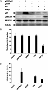
The activated NF-κB pathway alone is not sufficient for TGF-β1-mediated downregulation of nitric oxide production. (A) DG75 cells were transiently transfected with 5 μg of the luciferase reporter plasmid pNF-κB-Luc and 100 ng of internal control plasmid pRL-TK or with a combination of expression plasmids for p65 (pEGFP-p65), NIK (positive control), or an empty vector (pEGFP) or were treated (or not) with TGF-β1 (2 ng/ml). Cells were harvested and lysed, and equal amounts of protein separated by SDS-PAGE were then Western blotted with antibodies against p65, phospho-ERK 1/2, ERK 1/2, and tubulin. (B) The production of nitric oxide was measured by a nitrate/nitrite assay (Cayman). (C) NF-κB-dependent luciferase activity (luminescence) was measured using a dual-luciferase assay system (Promega, Madison, WI); relative light units (RLU) were calculated by normalizing firefly luciferase activity against Renilla luciferase activity.
Fig. 8.
Fig. 8.
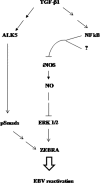
Schematic description of the TGF-β1-mediated pathway that leads to EBV reactivation in BL cells. First there is a quick and transient activation of NF-κB, and then there is a downregulation of iNOS activity that requires the participation of NF-κB, leading to ERK 1/2 phosphorylation. Although necessary, NF-κB alone is not sufficient to produce downregulation of iNOS, suggesting that another, uncharacterized event(s) is involved at this step. Simultaneously, TGF-β1 mediates Smad pathway activation through its receptor I/ALK5. However, the activated canonical pathway (involving Smad factors) and noncanonical pathways (involving NF-κB, iNOS, and ERK 1/2) in concert contribute to TGF-β1-mediated ZEBRA expression and consequently EBV reactivation in BL cells.

Similar articles

Cited by

References

    1. Agresti A., Lupo R., Bianchi M. E., Muller S. 2003. HMGB1 interacts differentially with members of the Rel family of transcription factors. Biochem. Biophys. Res. Commun. 302:421–426 - PubMed
    1. Arbach H., et al. 2006. Epstein-Barr virus (EBV) genome and expression in breast cancer tissue: effect of EBV infection of breast cancer cells on resistance to paclitaxel (Taxol). J. Virol. 80:845–853 - PMC - PubMed
    1. Arsura M., et al. 2003. Transient activation of NF-kappaB through a TAK1/IKK kinase pathway by TGF-beta1 inhibits AP-1/SMAD signaling and apoptosis: implications in liver tumor formation. Oncogene 22:412–425 - PubMed
    1. Bierie B., Moses H. L. 2006. Tumour microenvironment: TGFbeta: the molecular Jekyll and Hyde of cancer. Nat. Rev. Cancer 6:506–520 - PubMed
    1. Bonavida B., Baritaki S. 2011. Dual role of NO donors in the reversal of tumor cell resistance and EMT: Downregulation of the NF-kappaB/Snail/YY1/RKIP circuitry. Nitric Oxide 24:1–11 - PubMed

Publication types

MeSH terms

LinkOut - more resources