. 2011 Jul;122(1):26-37.
doi: 10.1093/toxsci/kfr089. Epub 2011 Apr 19.

Detoxification: a novel function of BRCA1 in tumor suppression?

Affiliations

Detoxification: a novel function of BRCA1 in tumor suppression?

Hyo Jin Kang et al. Toxicol Sci. 2011 Jul.

Abstract

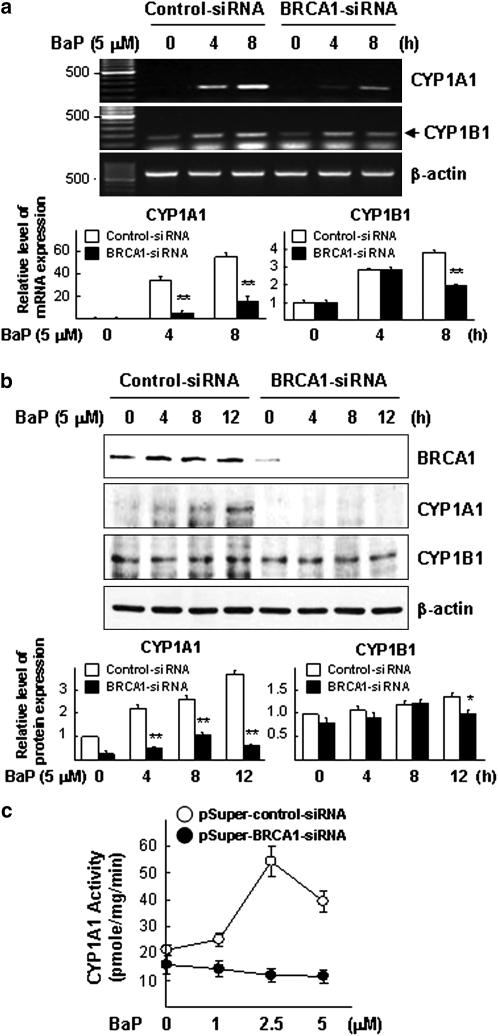
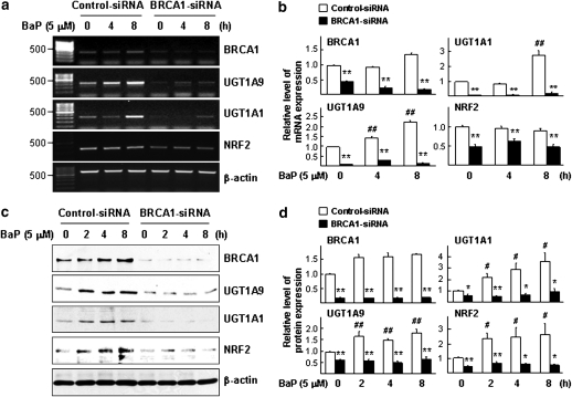
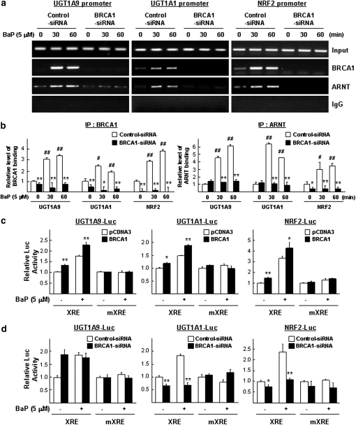
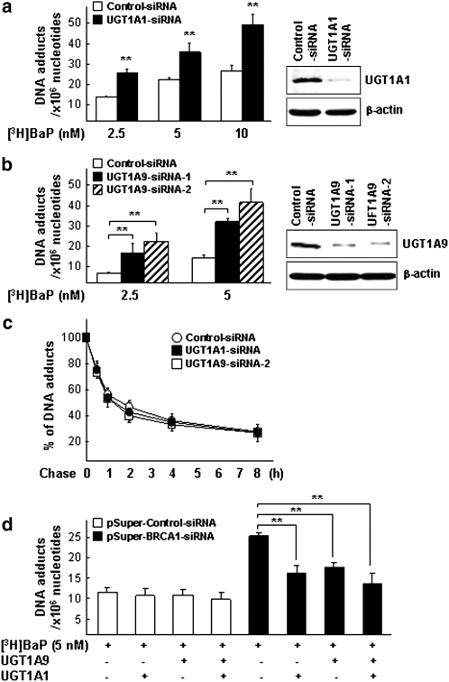
Our studies found that BRCA1 levels negatively correlate with DNA adducts induced by Benzo(a)pyrene (BaP). Pulse-chase experiments showed that the increase in BaP-induced DNA adducts in BRCA1 knockdown cells may not be associated with BRCA1's function in nucleotide excision repair activity; rather, it may be associated with its function in modulating transcriptional regulation. BRCA1 knockdown in MCF-10A cells significantly attenuated the induction of CYP1A1 following BaP treatment indicating that the increase in BaP-induced adducts in BRCA1 knockdown cells is not CYP1A1 dependent. However, our study shows that BRCA1 defective cells may still be able to biotransform BaP by regulating other CYP enzymes, including CYP1B1. Knockdown of BRCA1 also severely affected the expression levels of two types of uridine diphosphate glucorunyltransferase (UGT1A1 and UGT1A9) and NRF2. Both UGTs are known as BaP-specific detoxification enzymes, and NRF2 is a master regulator of antioxidant and detoxification genes. Thus, we concluded that the increased amount of BaP-induced DNA adducts in BRCA1 knockdown cells is strongly associated with its loss of functional detoxification. Chromatin immunoprecipitation assay revealed that BRCA1 is recruited to the promoter/enhancer sequences of UGT1A1, UGT1A9, and NRF2. Regulation of UGT1A1 and UGT1A9 expression showed that the induction of DNA adducts by BaP is directly affected by their expression levels. Finally, overexpression of UGTs, NRF2, or ARNT significantly decreased the amount of BaP-induced adducts in BRCA1-deficient cells. Overall, our results suggest that BRCA1 protects cells by reducing the amount of BaP-induced DNA adducts possibly via transcriptional activation of detoxification gene expression.


Figures

FIG. 1.
FIG. 1.
Overexpression of BRCA1 decreases the amount of DNA adducts induced by BaP. (a) wt BRCA1 overexpression decreases the amount of DNA adducts induced by [3H]BaP in human BRCA1 mutated breast cancer cell lines, SUM149PT and SUM1315MO2, as well as the nearly normal-like human breast cell line, MCF-10A. All three breast cell lines were transfected with expression vectors (pCDNA3 vs. pCDNA3-BRCA1) overnight and were cultured for an additional 24 h in 2.5 or 5nM of [3H]BaP. Analysis by WB demonstrated wt BRCA1 overexpression in MCF-10A cells. (b) Exogenously expressed wt BRCA1 or mutant BRCA1(s) differentially affect(s) the amount of DNA adducts induced by BaP in SUM149PT cells. Cells transfected with various BRCA1 expression vectors were treated with 5nM of [3H]BaP for 24 h, and radioactivity levels were determined. Analysis by WB was done using an anti-BRCA1 antibody to assure that the differential effects on the amount of BaP-induced adducts were not due to differential BRCA1 expression levels. β-Actin was used as a loading control. Student’s t-tests were applied for statistical significance; (*) is p < 0.05 and (**) indicates p < 0.01.
FIG. 2.
FIG. 2.
BRCA1 knockdown or knockout increases the amount of DNA adducts induced by carcinogens (BaP or DMBA). (a) BRCA1 knockdown increases the amount of DNA adducts induced by [3H]BaP in MCF-10A cells. Cells were transfected with shRNA coding vector (pSuper-control vs. pSuper-BRCA1) for 72 h and then treated with 2.5 or 5 nM of [3H]BaP for an additional 24 h. (b) The effect of BRCA1 knockdown on the amount of adducts induced by BaP was measured by the [32P]post labeling method. MCF-10A cells transfected with DNA vectors containing shRNA for 72 h were treated with 5 μM of BaP for 24 h. Genomic DNA was isolated, [32P]post labeled, and analyzed by a TLC. BPDE was used as a positive control. (c). The effect of BRCA1 knockdown on the amount of DMBA-induced adducts. A similar experiment as in (b) was performed except with DMBA. The radioactivity of BaP- or DMBA-induced DNA adduct spots from BRCA1 knockdown cells was measured by scintillation counter and compared with controls as mean of relative radioactivity ± S.E. (d) The increase in DNA adducts induced by [3H]BaP in cultured whole mammary glands of Brca1 conditional knockout mice (co/co). The third mammary glands from BRCA1wt/wt (wt/wt) and BRCA1co/co (co/co) mice were treated with 5 nM of [3H]BaP for 24 h and then assayed to measure the amount of DNA adducts. The data presented in (a) and (d) are as the mean of the number of DNA adducts per 106 nucleotides ± S.E of three independent experiments. Student’s t-tests were applied for statistical significance; (*) is p < 0.05 and (**) indicates p < 0.01.
FIG. 3.
FIG. 3.
Kinetics of BaP-DNA adducts removal in BRCA1 and NER knockdown cells. (a) Pulse-chase experiments were used to monitor the effect of BRCA1 status on the removal of DNA adducts induced by [3H]BaP. MCF-10A cells were transfected with the shRNA containing vector for 72 h and treated with 5nM of [3H]BaP for 24 h. Cells were then washed and incubated in fresh media for the indicated times and the remaining levels of [3H]BaP adducts were measured. (b–d) Pulse-chase experiments were performed as in (a) to monitor the effect of XPA (b), ERCC1 (c), or XPA + ERCC1 (d) knockdowns (using chemical siRNA) on the removal of [3H]BaP-induced DNA adducts. (e) Knockdowns of each gene (XPA or ERCC1) were confirmed by WB analysis. Student’s t-tests were applied for statistical significance; (*) and (**) indicate p < 0.05 and p < 0.01, respectively.
FIG. 4.
FIG. 4.
Effect of endogenous BRCA1 on CYP1A1 and CYP1B1 gene, protein, and EROD activity. MCF-10A cells were pretreated with siRNA (control vs. BRCA1), treated with BaP and harvested for (a) CYP1A1 and CYP1B1 mRNA, (b) CYP1A1 and CYP1B1 protein, and (c) CYP1A1 enzymatic activity. Bar graphs show the results of semiquantitative RT-PCR (a) and WB (b) results of three independent experiments quantified by densitometry. Student’s t-tests were applied for statistical significance; (*) is p < 0.05 and (**) indicates p < 0.01.
FIG. 5.
FIG. 5.
BRCA1 regulates the expression of endogenous UGT1A1, UGT1A9, and NRF2. Differential gene expression of UGT1A1, UGT1A9, or NRF2 in the presence and absence of endogenous BRCA1 was determined by semiquantitative RT-PCR and WB analysis. (a) BRCA1 levels affect the mRNA level of UGT1A1, UGT1A9, and NRF2 following BaP. Cells were pretreated with siRNA (control vs. BRCA1) for 72 h, treated with 5μM of BaP, and harvested at the indicated time points. The agarose gel image is a representative of three independent experiments. (b) Mean ± SE values from three independent semiquantitative RT-PCR assays were plotted to compare relative level of each transcript. (c) Cells were pretreated with siRNA (control vs. BRCA1) for 72 h, treated with 5μM of BaP (0, 2, 4, or 8 h), and harvested for WB analysis. (d) Mean ± SE values from three independent WB analysis were plotted to show relative intensity values of each band as determined by densitometry. Student’s t-tests were applied for statistical significance. Symbols (*) and (**) indicate p < 0.05 and p < 0.01 (control- vs. BRCA1-siRNA in the same concentration of BaP), respectively; Symbols (#) and (##) indicate p < 0.05 and p < 0.01 (no treatment vs. BaP treatment in control-siRNA), respectively.
FIG. 6.
FIG. 6.
BRCA1 is recruited to UGT1A1, UGT1A9, and NRF2 promoter/enhancer sequences. (a) ChIP assays were performed to determine the effect of BRCA1 knockdown on promoter occupancy by UGT1A1, UGT1A9, and NRF2. Fragmented genomic DNA from MCF-10A cells, which had been pretreated with siRNA for 72 h and then treated with 5μM BaP for 30 or 60 min, was immunoprecipitated by anti-BRCA1 and anti-ARNT antibodies. (b) RT-PCR of (a) results. Student’s t-tests were applied for statistical significance; (#) and (##) indicate p < 0.05 and p < 0.01 (no treatment vs. BaP treatment in control-siRNA), respectively and (*) p < 0.05 and (**) p < 0.01 (control-siRNA vs. BRCA1-siRNA), respectively. (c) The effects of BRCA1 overexpression on BaP-induced UGT1A1, UGT1A9, and NRF2 promoter reporter activity (XRE and mutant XRE [mXRE]). Cells were transfected with the reporter gene and expression vector and treated with either DMSO or BaP for 24 h. (d) Effects of BRCA1 knockdown on BaP-induced reporter activity of UGT1A1, UGT1A9, and NRF2 promoter. Cells were pretreated with siRNA for 48 h, transfected with a reporter plasmid, and then treated with 5μM BaP for 24 h. Student’s t-tests were applied for statistical significance; (*) and (**) indicate p < 0.05 and p < 0.01 (control-siRNA vs. BRCA1-siRNA), respectively.
FIG. 7.
FIG. 7.
UGT1A1 or UGT1A9 regulates the amount of DNA adducts induced by BaP. (a, b) Effect of UGT1A1- or UGT1A9-siRNA on the amount of BaP-induced adducts. Cells were pretreated with siRNAs for UGT1A1 (a) or UGT1A9 (b) for 72 h and treated with 2.5, 5, or 10nM of [3H]BaP for 24 h. Knockdown of UGTs was confirmed by WB analysis (right panels). (c) Kinetics of removal of DNA adducts induced by [3H]BaP in UGT1A1 or UGT1A9 knockdown cells. Pulse-chase experiments were performed to determine effects of UGT1A1 or UGT1A9 knockdown on the removal of [3H]BaP-DNA adducts. (d) Restoring UGT1A1 and/or UGT1A9 genes decreased the amount of BaP-induced DNA adducts in BRCA1 knockdown MCF-10A cells. Cells transfected with shRNA-coding vector (control vs. BRCA1) for 48 h were transfected with 1 μg of expression vectors for either UGT1A1 or UGT1A9 or both for an additional 24 h. The transfected cells were further treated with 5nM of [3H]BaP for 24 h, and the amount of DNA adducts was measured. Student’s t-tests were applied for statistical significance; (**) indicates p < 0.01 (a, b, and d).
FIG. 8.
FIG. 8.
Effects of restoring ARNT or NRF2 on BaP-induced DNA adducts in BRCA1 knockdown cells. (a) Cells were pretreated with siRNA (control vs. BRCA1), treated with BaP (0, 2.5, and 5μM), harvested, and confirmed by WB analysis using anti-BRCA1, anti-NRF2, and anti-ARNT antibodies. (b) Overexpression of ARNT in BRCA1 knockdown cells decreases the amount of adducts induced by BaP. Cells transfected with shRNA coding vector (pSuper-control vs. pSuper-BRCA1) for 48 h were transfected with GFP-tagged ARNT (vs. pEGFP as a empty vector) and then treated with [3H]BaP for 24 h. (c) Restoring NRF2 reduced the amount of DNA adducts in BRCA1 knockdown cells. Cells were transfected with shRNA as in (a) and infected with adenovirus (Ad)-flag-NRF2 (vs. Ad-Null) and then [3H]BaP-DNA adducts were measured. Student’s t-tests were applied for statistical significance; (*) and (**) indicate p < 0.05 and p < 0.01, respectively. Far right panels of (b) and (c) show the results of WB analysis after restoring either GFP-ARNT or Ad-flag-NRF2 in BRCA1 knockdown cells.

Similar articles

Cited by

References

    1. Abbott DW, Thompson ME, Robinson-Benion C, Tomlinson G, Jensen RA, Holt JT. BRCA1 expression restores radiation resistance in BRCA1-defective cancer cells through enhancement of transcription-coupled DNA repair. J. Biol. Chem. 1999;274:18808–18812. - PubMed
    1. Ali AB, Iau PT, Sng JH. Cancer-specific methylation in the BRCA1 promoter in sporadic breast tumours. Med. Oncol. 2011;28:64–66. - PubMed
    1. Bae I, Fan S, Meng Q, Rih JK, Kim HJ, Kang HJ, Xu J, Goldberg ID, Jaiswal AK, Rosen EM. BRCA1 induces antioxidant gene expression and resistance to oxidative stress. Cancer Res. 2004;64:7893–7909. - PubMed
    1. Bochar DA, Wang L, Beniya H, Kinev A, Xue Y, Lane WS, Wang W, Kashanchi F, Shiekhattar R. BRCA1 is associated with a human SWI/SNF-related complex: linking chromatin remodeling to breast cancer. Cell. 2000;102:257–265. - PubMed
    1. Breast Cancer Family Registry, Kathleen Cuningham Consortium for Research into Familial Breast Cancer (Australasia), Ontario Cancer Genetics Network (Canada) Smoking and risk of breast cancer in carriers of mutations in BRCA1 or BRCA2 aged less than 50 years. Breast Cancer Res. Treat. 2008;109:67–75. - PMC - PubMed

Publication types

MeSH terms