

Notch signaling modulates MUC16 biosynthesis in an in vitro model of human corneal and conjunctival epithelial cell differentiation
- PMID: 21508102
- PMCID: PMC3176047
- DOI: 10.1167/iovs.11-7196
Notch signaling modulates MUC16 biosynthesis in an in vitro model of human corneal and conjunctival epithelial cell differentiation
Abstract
Purpose: Notch proteins are a family of transmembrane receptors that coordinate binary cell fate decisions and differentiation in wet-surfaced epithelia. We sought to determine whether Notch signaling contributes to maintaining mucosal homeostasis by modulating the biosynthesis of cell surface-associated mucins in an in vitro model of human corneal (HCLE) and conjunctival (HCjE) epithelial cell differentiation.
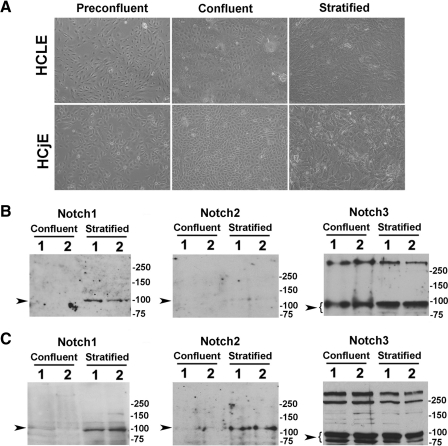
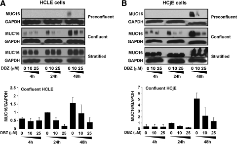
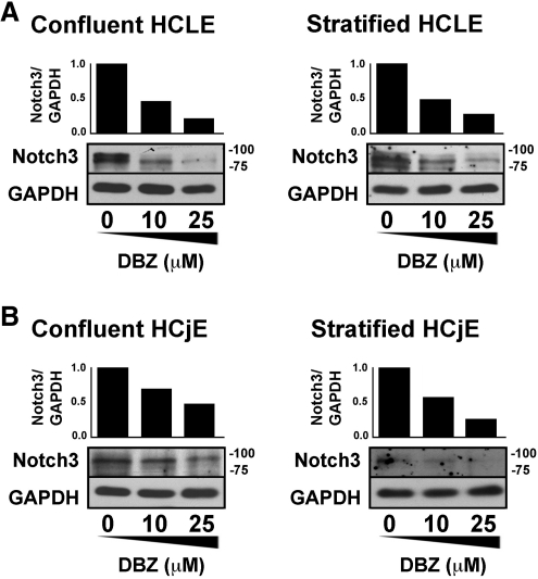
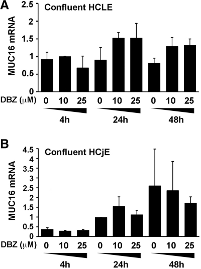
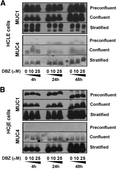
Methods: HCLE and HCjE cells were grown at different stages of differentiation, representing nondifferentiated (preconfluent and confluent) and differentiated (stratified) epithelial cultures. Notch signaling was blocked with the γ-secretase inhibitor dibenzazepine (DBZ). The presence of Notch intracellular domains (Notch1 to Notch3) and mucin protein (MUC1, -4, -16) was evaluated by electrophoresis and Western blot analysis. Mucin gene expression was determined by TaqMan real-time polymerase chain reaction.
Results: Here we demonstrate that Notch3 is highly expressed in undifferentiated and differentiated HCLE and HCjE cells, and that Notch1 and Notch2 biosynthesis is enhanced by induction of differentiation with serum-containing media. Inhibition of Notch signaling with DBZ impaired MUC16 biosynthesis in a concentration-dependent manner in undifferentiated cells at both preconfluent and confluent stages, but not in postmitotic stratified cells. In contrast to protein levels, the amount of MUC16 transcripts were not significantly reduced after DBZ treatment, suggesting that Notch regulates MUC16 posttranscriptionally. Immunoblots of DBZ-treated epithelial cells grown at different stages of differentiation revealed no differences in the levels of MUC1 and MUC4.
Conclusions: These results indicate that MUC16 biosynthesis is posttranscriptionally regulated by Notch signaling at early stages of epithelial cell differentiation, and suggest that Notch activation contributes to maintaining a mucosal phenotype at the ocular surface.
Figures
Similar articles
-
Differential effect of rebamipide on transmembrane mucin biosynthesis in stratified ocular surface epithelial cells.Exp Eye Res. 2016 Dec;153:1-7. doi: 10.1016/j.exer.2016.10.007. Epub 2016 Oct 8. Exp Eye Res. 2016. PMID: 27725198
-
MUC16 mucin is expressed by the human ocular surface epithelia and carries the H185 carbohydrate epitope.Invest Ophthalmol Vis Sci. 2003 Jun;44(6):2487-95. doi: 10.1167/iovs.02-0862. Invest Ophthalmol Vis Sci. 2003. PMID: 12766047
-
Differential regulation of membrane-associated mucins in the human ocular surface epithelium.Invest Ophthalmol Vis Sci. 2004 Jan;45(1):114-22. doi: 10.1167/iovs.03-0903. Invest Ophthalmol Vis Sci. 2004. PMID: 14691162
-
Membrane-tethered mucins have multiple functions on the ocular surface.Exp Eye Res. 2010 Jun;90(6):655-63. doi: 10.1016/j.exer.2010.02.014. Epub 2010 Mar 16. Exp Eye Res. 2010. PMID: 20223235 Free PMC article. Review.
-
Functions of ocular surface mucins in health and disease.Curr Opin Allergy Clin Immunol. 2008 Oct;8(5):477-83. doi: 10.1097/ACI.0b013e32830e6b04. Curr Opin Allergy Clin Immunol. 2008. PMID: 18769205 Free PMC article. Review.
Cited by
-
Primary cilia maintain corneal epithelial homeostasis by regulation of the Notch signaling pathway.Development. 2016 Jun 15;143(12):2160-71. doi: 10.1242/dev.132704. Epub 2016 Apr 27. Development. 2016. PMID: 27122169 Free PMC article.
-
Tear film mucins: front line defenders of the ocular surface; comparison with airway and gastrointestinal tract mucins.Exp Eye Res. 2013 Dec;117:62-78. doi: 10.1016/j.exer.2013.07.027. Epub 2013 Aug 14. Exp Eye Res. 2013. PMID: 23954166 Free PMC article.
-
Expression analysis of the transmembrane mucin MUC20 in human corneal and conjunctival epithelia.Invest Ophthalmol Vis Sci. 2014 Aug 28;55(10):6132-8. doi: 10.1167/iovs.14-15269. Invest Ophthalmol Vis Sci. 2014. PMID: 25168902 Free PMC article.
-
A novel fluorescent lipid probe for dry eye: retrieval by tear lipocalin in humans.Invest Ophthalmol Vis Sci. 2013 Feb 19;54(2):1398-410. doi: 10.1167/iovs.12-10817. Invest Ophthalmol Vis Sci. 2013. PMID: 23361507 Free PMC article.
-
The ocular surface epithelial barrier and other mechanisms of mucosal protection: from allergy to infectious diseases.Curr Opin Allergy Clin Immunol. 2013 Oct;13(5):563-8. doi: 10.1097/ACI.0b013e3283645899. Curr Opin Allergy Clin Immunol. 2013. PMID: 23974687 Free PMC article. Review.
References
-
- Fortini ME. Notch signaling: the core pathway and its posttranslational regulation. Dev Cell. 2009;16:633–647 - PubMed
-
- van Es JH, van Gijn ME, Riccio O, et al. Notch/gamma-secretase inhibition turns proliferative cells in intestinal crypts and adenomas into goblet cells. Nature. 2005;435:959–963 - PubMed
-
- Nicolas M, Wolfer A, Raj K, et al. Notch1 functions as a tumor suppressor in mouse skin. Nat Genet. 2003;33:416–421 - PubMed
-
- Ma A, Boulton M, Zhao B, Connon C, Cai J, Albon J. A role for Notch signaling in human corneal epithelial cell differentiation and proliferation. Invest Ophthalmol Vis Sci. 2007;48:3576–3585 - PubMed
Publication types
MeSH terms
Substances
Grants and funding
LinkOut - more resources
Full Text Sources
Research Materials
Miscellaneous
