

Polycystin-2 expression and function in adult mouse lacrimal acinar cells
- PMID: 21508103
- PMCID: PMC3176037
- DOI: 10.1167/iovs.10-7114
Polycystin-2 expression and function in adult mouse lacrimal acinar cells
Abstract
Purpose: Lacrimal glands regulate the production and secretion of tear fluid. Dysfunction of lacrimal gland acinar cells can ultimately result in ocular surface disorders, such as dry eye disease. Ca(2+) homeostasis is tightly regulated in the cellular environment, and secretion from the acinar cells of the lacrimal gland is regulated by both cholinergic and adrenergic stimuli, which both result in changes in the cytosolic Ca(2+) concentration. We have previously described the detailed intracellular distribution of inositol-1,4,5-trisphosphate receptors (IP(3)Rs), and ryanodine receptors (RyRs) in lacrimal acinar cells, however, little is known regarding the expression and distribution of the third major class of intracellular Ca(2+) release channels, transient receptor potential polycystin family (TRPP) channels.
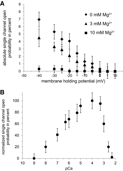
Methods: Studies were performed in adult lacrimal gland tissue of Swiss-Webster mice. Expression, localization, and intracellular distribution of TRPP Ca(2+) channels were investigated using immunocytochemistry, immunohistochemistry, and electron microscopy. The biophysical properties of single polycystin-2 channels were investigated using a planar lipid bilayer electrophysiology system.
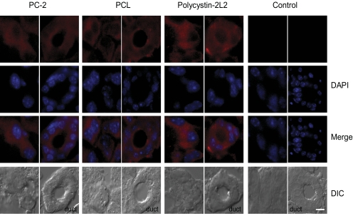
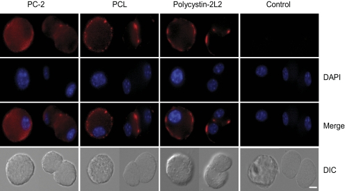
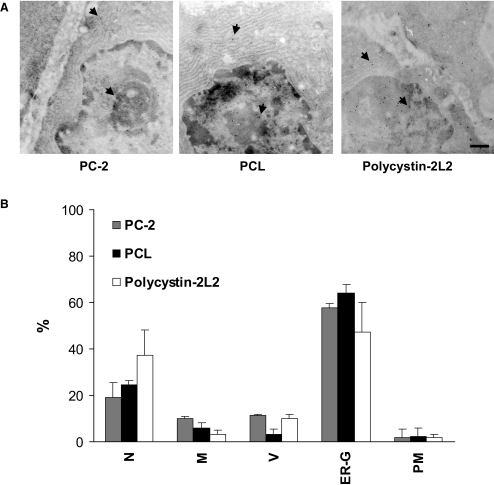
Results: All channel-forming isoforms of TRPP channels (polycystin-2, polycystin-L, and polycystin-2L2) were expressed in adult mouse lacrimal gland. Subcellular analysis of immunogold labeling revealed strongest polycystin-2 expression on the membranes of the endoplasmic reticulum, Golgi, and nucleus. Biophysical properties of lacrimal gland polycystin-2 channels were similar to those described for other tissues.
Conclusions: The expression of TRPP channels in lacrimal acinar cells suggests a functional role of the proteins in the regulation of lacrimal fluid secretion under physiological and disease conditions, and provides the basis for future studies focusing on physiology and pharmacology.
Figures
References
-
- Bromberg BB. Autonomic control of lacrimal protein secretion. Invest Ophthalmol Vis Sci. 1981;20:110–116 - PubMed
-
- Sundermeier T, Matthews G, Brink PR, Walcott B. Calcium dependence of exocytosis in lacrimal gland acinar cells. Am J Physiol Cell Physiol. 2002;282:C360–C365 - PubMed
-
- Yu J, Asche CV, Fairchild CJ. The economic burden of dry eye disease in the United States: a decision tree analysis. Cornea. 2010;30:379–387 - PubMed
-
- Koulen P, Thrower EC. Pharmacological modulation of intracellular Ca(2+) channels at the single-channel level. Mol Neurobiol. 2001;24:65–86 - PubMed
Publication types
MeSH terms
Substances
Grants and funding
- P01 AG022550/AG/NIA NIH HHS/United States
- EY015672/EY/NEI NIH HHS/United States
- P01 AG027956/AG/NIA NIH HHS/United States
- AG022550/AG/NIA NIH HHS/United States
- P01 AG010485/AG/NIA NIH HHS/United States
- EY014227/EY/NEI NIH HHS/United States
- R21 EY015672/EY/NEI NIH HHS/United States
- RR022570/RR/NCRR NIH HHS/United States
- RR027093/RR/NCRR NIH HHS/United States
- AG010485/AG/NIA NIH HHS/United States
- R01 EY014227/EY/NEI NIH HHS/United States
- AG027956/AG/NIA NIH HHS/United States
- S10 RR027093/RR/NCRR NIH HHS/United States
- S10 RR022570/RR/NCRR NIH HHS/United States
LinkOut - more resources
Full Text Sources
Medical
Molecular Biology Databases
Miscellaneous
