. 2011 Apr 20;31(16):6019-27.
doi: 10.1523/JNEUROSCI.6498-10.2011.

Nutrient-sensing hypothalamic TXNIP links nutrient excess to energy imbalance in mice

Affiliations

Nutrient-sensing hypothalamic TXNIP links nutrient excess to energy imbalance in mice

Clémence Blouet et al. J Neurosci. .

Abstract

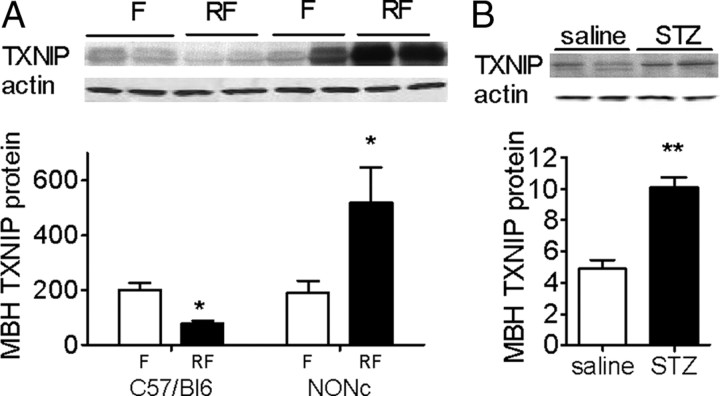
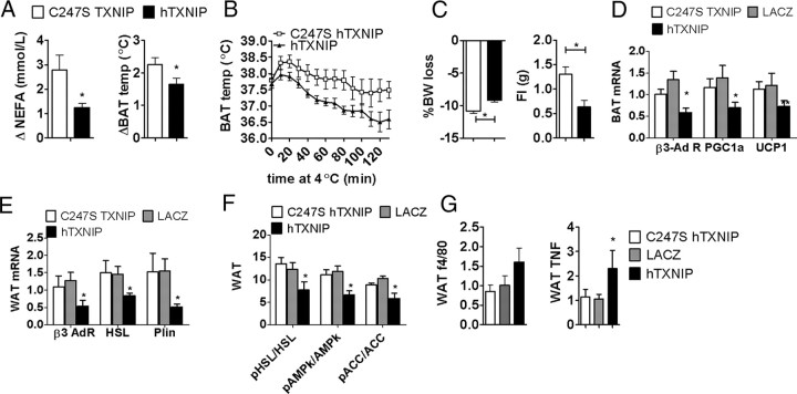
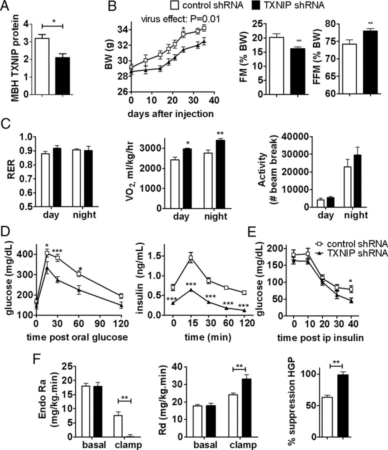
Nutrient excess in obesity and diabetes is emerging as a common putative cause for multiple deleterious effects across diverse cell types, responsible for a variety of metabolic dysfunctions. The hypothalamus is acknowledged as an important regulator of whole-body energy homeostasis, through both detection of nutrient availability and coordination of effectors that determine nutrient intake and utilization, thus preventing cellular and whole-body nutrient excess. However, the mechanisms underlying hypothalamic nutrient detection and its impact on peripheral nutrient utilization remain poorly understood. Recent data suggest a role for thioredoxin-interacting protein (TXNIP) as a molecular nutrient sensor important in the regulation of energy metabolism, but the role of hypothalamic TXNIP in the regulation of energy balance has not been evaluated. Here we show in mice that TXNIP is expressed in nutrient-sensing neurons of the mediobasal hypothalamus, responds to hormonal and nutrient signals, and regulates adipose tissue metabolism, fuel partitioning, and glucose homeostasis. Hypothalamic expression of TXNIP is induced by acute nutrient excess and in mouse models of obesity and diabetes, and downregulation of mediobasal hypothalamic TXNIP expression prevents diet-induced obesity and insulin resistance. Thus, mediobasal hypothalamic TXNIP plays a critical role in nutrient sensing and the regulation of fuel utilization.


Figures

Figure 1.
Figure 1.
TXNIP is expressed in the hypothalamus and is suppressed by nutritional and hormonal signals of energy availability. Immunohistochemistry showing TXNIP in the MBH and the PVN of a C3H wild-type control (A) and a Hcb-19 TXNIP-deficient mouse (B). C, Immunohistochemistry showing TXNIP expression in the arcuate (Arc), the ventromedial (VMH), the PVN, and the LH nuclei of the hypothalamus in a C57BL/6 mouse. D, Western blot analyses of MBH TXNIP expression in C3H and Hcb-19 mice. E, TXNIP mRNA and protein expression corrected to actin, and thioredoxin activity in the MBH, PVN, and/or LH of 24 h fasted (F) and 4 h refed (RF) mice (n = 4–6). F, MBH TXNIP protein expression corrected to actin after an acute intra-MBH infusion of aCSF, leptin, or insulin (n = 5). All data are means ± SEM. *p < 0.05, **p < 0.01 versus controls.
Figure 2.
Figure 2.
MBH TXNIP expression in mouse models of diabetes. MBH TXNIP protein expression corrected to actin in 24 h fasted (F) and 4 h refed (RF) NONcNZ10/LtJ mice (NONc; A) and streptozotocin (STZ)-treated mice (B). All data are means ± SEM. *p < 0.05, **p < 0.01, ***p < 0.001 versus controls.
Figure 3.
Figure 3.
MBH TXNIP overexpression increases body weight and body fat and decreases energy expenditure. A, TXNIP protein expression corrected to actin in the MBH, PVN, and LH of mice infected with hTXNIP (hT) or LacZ (LZ) lentivirus (n = 4), and viral infection spread (as shown by EGFP immunofluorescence) after MBH stereotaxic infection with an EGFP lentivirus. Body weight, daily food intake, fat mass (FM), and fat free mass (FFM) (n = 6–10) (B) and oxygen consumption, respiratory exchange ratio, total spontaneous locomotor, and brown fat temperature (n = 4) (C) in mice fed a high-fat diet after MBH injection of C247S hTXNIP, LacZ, or hTXNIP lentivirus. All data are means ± SEM. *p < 0.05, **p < 0.01 versus controls. ARC, Arcuate nucleus; DMN, dorsomedial hypothalamus; HFD, high-fat diet; RER, respiratory exchange ratio; BAT, brown adipose tissue.
Figure 4.
Figure 4.
MBH TXNIP overexpression impairs glycemic control and nutrient utilization. Mice were injected with the C247S hTXNIP, LacZ, or hTXNIP lentivirus into the MBH, fed a high-fat diet after the viral injections, and studied in the 24 h fasted (RF) or 3 h fed (F) state between 4 and 6 weeks after the viral injection. Blood glucose and plasma insulin (n = 10) (A), blood glucose during an intraperitoneal insulin sensitivity test (n = 10) (B), blood glucose and plasma insulin during an oral glucose tolerance test (n = 10) (C), endo Ra, Rd, and percentage suppression of hepatic glucose production (HGP) during the basal and hyperinsulinemic (clamp) period of a euglycemic clamp (n = 4–5) (D), and plasma triglycerides (TG), plasma β-hydroxybutyrate (β-HB), and plasma non-esterified free fatty acids (n = 10) (E). All data are means ± SEM. *p < 0.05, **p < 0.01 versus controls.
Figure 5.
Figure 5.
Increased adiposity during MBH TXNIP overexpression contributes to the impairment in glycemic control. Body weight, fat mass (FM), fat free mass (FFM), blood glucose during an oral glucose tolerance test (OGTT), and an intraperitoneal insulin sensitivity test (IST) in hTXNIP-expressing mice fed ad libitum or restricted to match the body weight of C247S hTXNIP-expressing controls for 4 weeks. All data are means ± SEM; n = 6. *p < 0.05, **p < 0.01 versus controls.
Figure 6.
Figure 6.
MBH TXNIP overexpression impairs sympathetic activity to white and brown fat and adipose tissue metabolism. Plasma NEFA and brown fat temperature changes after an intraperitoneal administration of CL316243, a β3 receptor agonist (A), cold sensitivity during a 2 h cold challenge at 4°C (B), body weight loss during a 24 h fast and food intake during the subsequent 4 h refeeding in mice injected with C247S or hTXNIP lentivirus into the MBH (n = 5) (C). Brown fat PGC1α, UCP1, and β3 adrenergic receptor mRNA expression corrected to actin (D), visceral fat β3 adrenergic receptor, HSL, and perilipin (Plin) mRNA expression corrected to actin (E), visceral fat AMPkα Thr172 phosphorylation and HSL Ser 563 phosphorylation (F), and visceral fat F4/80 and TNF mRNA expression corrected to actin in fasted mice injected with C247S hTXNIP, LacZ, or hTXNIP lentivirus into the MBH (G). All data are means ± SEM. *p < 0.05, **p < 0.01 versus controls. BAT, Brown adipose tissue; WAT, white adipose tissue.
Figure 7.
Figure 7.
TXNIP overexpression in the MBH impairs MBH leptin action. A, Mediobasal hypothalamic leptin-induced STAT3 Y705 phosphorylation in mice expressing hTXNIP or C247S hTXNIP in the MBH (n = 5). B, Leptin-induced anorexia and body weight change in mice expressing hTXNIP or C247S hTXNIP in the MBH after an intra-MBH leptin (L; 150 ng) or aCSF (A) injection (n = 4). All data are means ± SEM. *p < 0.05, **p < 0.01, ***p < 0.001 versus controls.
Figure 8.
Figure 8.
Downregulation of MBH TXNIP expression protects from diet-induced obesity and insulin resistance. TXNIP protein expression corrected to actin in MBH of mice infected with TXNIP or control shRNA lentiviruses (n = 4) (A), body weight, fat mass (FM), fat free mass (FFM), and food intake (B), respiratory quotient, oxygen consumption, and locomotor activity (C), blood glucose and plasma insulin during an oral glucose tolerance test (D), blood glucose during an intraperitoneal insulin sensitivity test (E), and endo Ra, Rd, and percentage suppression of hepatic glucose production (HGP) during a euglycemic hyperinsulinemic clamp (D) in HFD-fed mice expressing TXNIP shRNA or a control shRNA in the MBH (n = 5–8) (F). All values are means ± SEM. *p < 0.05, **p < 0.01 versus control.

Similar articles

Cited by

References

    1. Andrews ZB, Diano S, Horvath TL. Mitochondrial uncoupling proteins in the CNS: in support of function and survival. Nat Rev Neurosci. 2005;6:829–840. - PubMed
    1. Azzara AV, Sokolnicki JP, Schwartz GJ. Central melanocortin receptor agonist reduces spontaneous and scheduled meal size but does not augment duodenal preload-induced feeding inhibition. Physiol Behav. 2002;77:411–416. - PubMed
    1. Benani A, Troy S, Carmona MC, Fioramonti X, Lorsignol A, Leloup C, Casteilla L, Pénicaud L. Role for mitochondrial reactive oxygen species in brain lipid sensing: redox regulation of food intake. Diabetes. 2007;56:152–160. - PubMed
    1. Bence KK, Delibegovic M, Xue B, Gorgun CZ, Hotamisligil GS, Neel BG, Kahn BB. Neuronal PTP1B regulates body weight, adiposity and leptin action. Nat Med. 2006;12:917–924. - PubMed
    1. Blouet C, Ono H, Schwartz GJ. Mediobasal hypothalamic p70 S6 kinase 1 modulates the control of energy homeostasis. Cell Metab. 2008;8:459–467. - PMC - PubMed

Publication types

MeSH terms

LinkOut - more resources