. 2011 Apr 20;31(16):6053-8.
doi: 10.1523/JNEUROSCI.5524-09.2011.

Astrocytes from the contused spinal cord inhibit oligodendrocyte differentiation of adult oligodendrocyte precursor cells by increasing the expression of bone morphogenetic proteins

Affiliations

Astrocytes from the contused spinal cord inhibit oligodendrocyte differentiation of adult oligodendrocyte precursor cells by increasing the expression of bone morphogenetic proteins

Yaping Wang et al. J Neurosci. .

Abstract

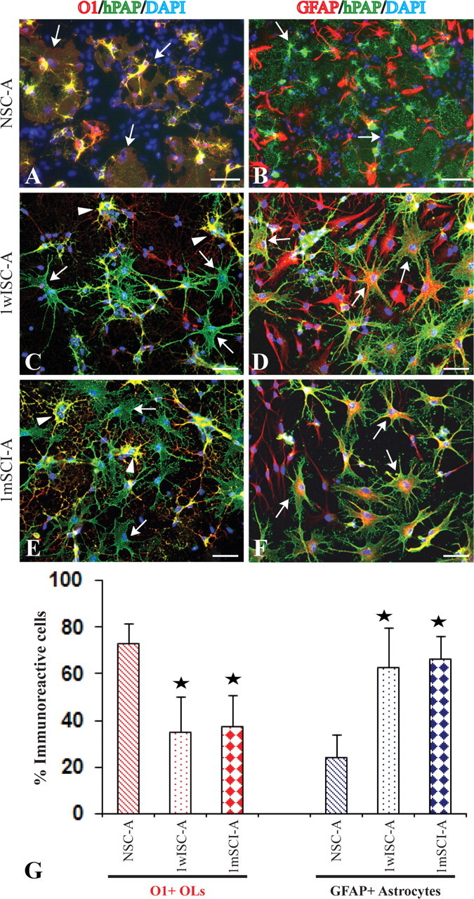
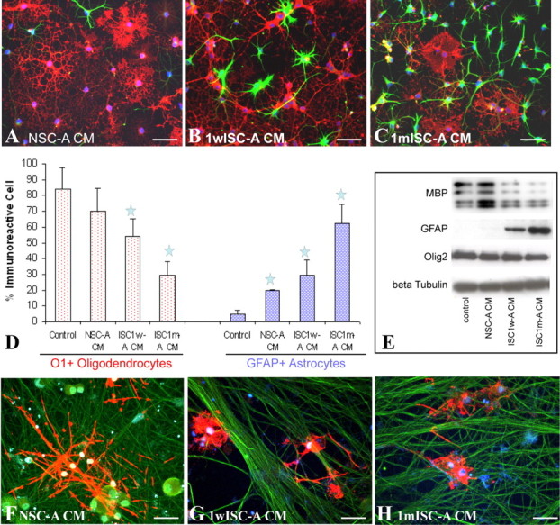
Promotion of remyelination is an important therapeutic strategy to facilitate functional recovery after traumatic spinal cord injury (SCI). Transplantation of neural stem cells (NSCs) or oligodendrocyte precursor cells (OPCs) has been used to enhance remyelination after SCI. However, the microenvironment in the injured spinal cord is inhibitory for oligodendrocyte (OL) differentiation of NSCs or OPCs. Identifying the signaling pathways that inhibit OL differentiation in the injured spinal cord could lead to new therapeutic strategies to enhance remyelination and functional recovery after SCI. In the present study, we show that reactive astrocytes from the injured rat spinal cord or their conditioned media inhibit OL differentiation of adult OPCs with concurrent promotion of astrocyte differentiation. The expression of bone morphogenetic proteins (BMP) is dramatically increased in the reactive astrocytes and their conditioned media. Importantly, blocking BMP activity by BMP receptor antagonist, noggin, reverse the effects of active astrocytes on OPC differentiation by increasing the differentiation of OL from OPCs while decreasing the generation of astrocytes. These data indicate that the upregulated bone morphogenetic proteins in the reactive astrocytes are major factors to inhibit OL differentiation of OPCs and to promote its astrocyte differentiation. These data suggest that manipulation of BMP signaling in the endogenous or grafted NSCs or OPCs may be a useful therapeutic strategy to increase their OL differentiation and remyelination and enhance functional recovery after SCI.


Figures

Figure 1.
Figure 1.
Characterization of OPCs and astrocytes. A–C, All purified OPCs express hPAP (A) and OPC markers O4 (A, B), olig2 (B), A2B5 (C), and Sox10 (C). D, E, OPCs differentiate into O1+ OLs after withdrawal of FGF2 and PDGF-aa for 3 d (D), and MBP+ OLs to form myelin around the axons after coculture with DRG neurons for 2 weeks (E). F, All purified astrocytes from 1 month postinjury spinal cord express GFAP. Some astrocytes are still proliferating as evidenced by BrdU incorporation. Scale bar, 50 μm.
Figure 2.
Figure 2.
Active astrocytes from the injured spinal cord inhibit OL differentiation of cocultured OPCs. Three days after being cocultured with astrocytes from normal spinal cord, the majority of OPCs labeled by hPAP differentiate into O1+ OLs with membrane sheets, the typical morphology of mature OLs (A, arrows). Most hPAP+ OPCs do not differentiate into GFAP+ astrocytes (B, arrowheads). However, in the coculture with astrocytes from 1 week (C, arrows) or 1 month (E, arrows) postinjury spinal cord, most OPCs labeled by hPAP fail to differentiate into O1+ OLs. OPCs, which differentiate into O1+ OLs, lack the complex membrane sheets (C, E, arrowheads). Most OPCs labeled by hPAP (green) differentiate into GFAP+ astrocytes (red) (D, F, arrows). Quantitative data confirm that reactive astrocytes from the injured spinal cord significantly decrease OL differentiation with a concurrent increase of astrocyte differentiation of cocultured OPCs (G). Data represent the mean ± SD from four repeated experiments from separately generated cultures; *p < 0.05. Scale bar, 50 μm.
Figure 3.
Figure 3.
Conditioned media from reactive astrocytes from the injured spinal cord inhibit OL differentiation of cocultured OPCs. Adult OPCs are differentiated for 3 d in control basal medium, NSC-A, 1wISC-A, or 1mISC-A. In basal medium or NSC-A CM, the majority of OPCs differentiate into O1+ OL (A, red) with few into GFAP+ astrocytes (A, green). However, in CM of ISC1w-A (B, D) or ISC1m-A (C, D), the number of OPCs that differentiated into OLs dramatically decreases while the number of GFAP+ astrocytes significantly increases. Western blot experiments confirm these immunohistochemical results (E). After coculture with DRG neurons for 12 d in CM from NSC-A, OPCs differentiate into MBP+ mature OLs, which form myelin along axons. However, myelin formation is not observed, although OPCs differentiate into MBP+ OLs in CM of 1wISC-A (G) or 1mISC-A (H). Data in D represent the mean ± SD from four independent experiments; *p < 0.05. Scale bar, 50 μm.
Figure 4.
Figure 4.
ISC1w-A and ISC1m-A inhibit OL differentiation of OPCs by increasing expression of BMPs. A, B, Expression of BMP2 and 4 is significantly increased in ISC1w- or ISC1m-A (A) or their CMs (B). C, CMs of 1wISC- or 1mISC-A increase the expression of pSMAD 1/5/8 in OPCs with decreasing expression of MBP and increasing expression of GFAP. Addition of noggin blocks the expression pSMAD and significantly increases and decreases the expression of MBP and GFAP, respectively (C). D–G, Immunohistochemistry further confirmed that blocking BMP signaling by noggin reverse the inhibition of the ISC1w or ISC1m-A CMs to significantly increase the number of OPCs differentiated into OLs with a concurrent decrease of astrocyte differentiation. OL or astrocyte differentiation in control basal medium or NSC-A CMs, however, was not changed by the addition of noggin. Data in F and G represent the mean ± SD from four repeated experiments; **p < 0.01. Scale bar, 50 μm.

Similar articles

Cited by

References

    1. Anderson JM, Hampton DW, Patani R, Pryce G, Crowther RA, Reynolds R, Franklin RJ, Giovannoni G, Compston DA, Baker D, Spillantini MG, Chandran S. Abnormally phosphorylated tau is associated with neuronal and axonal loss in experimental autoimmune encephalomyelitis and multiple sclerosis. Brain. 2008;131:1736–1748. - PubMed
    1. Ara J, See J, Mamontov P, Hahn A, Bannerman P, Pleasure D, Grinspan JB. Bone morphogenetic proteins 4, 6, and 7 are up-regulated in mouse spinal cord during experimental autoimmune encephalomyelitis. J Neurosci Res. 2008;86:125–135. - PubMed
    1. Back SA, Tuohy TM, Chen H, Wallingford N, Craig A, Struve J, Luo NL, Banine F, Liu Y, Chang A, Trapp BD, Bebo BF, Jr, Rao MS, Sherman LS. Hyaluronan accumulates in demyelinated lesions and inhibits oligodendrocyte progenitor maturation. Nat Med. 2005;11:966–972. - PubMed
    1. Blakemore WF, Gilson JM, Crang AJ. The presence of astrocytes in areas of demyelination influences remyelination following transplantation of oligodendrocyte progenitors. Exp Neurol. 2003;184:955–963. - PubMed
    1. Chang A, Tourtellotte WW, Rudick R, Trapp BD. Premyelinating oligodendrocytes in chronic lesions of multiple sclerosis. N Engl J Med. 2002;346:165–173. - PubMed

Publication types

MeSH terms

Substances