. 2011 Apr 20;31(16):6235-46.
doi: 10.1523/JNEUROSCI.5905-10.2011.

Oligodendrocyte progenitors reversibly exit the cell cycle and give rise to astrocytes in response to interferon-γ

Affiliations

Oligodendrocyte progenitors reversibly exit the cell cycle and give rise to astrocytes in response to interferon-γ

Daniel C Tanner et al. J Neurosci. .

Abstract

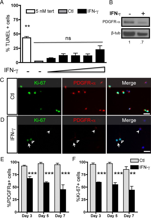
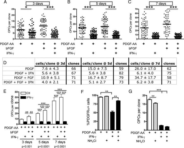
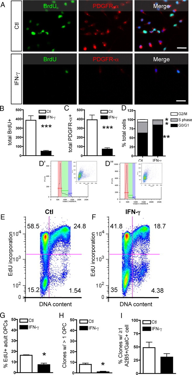
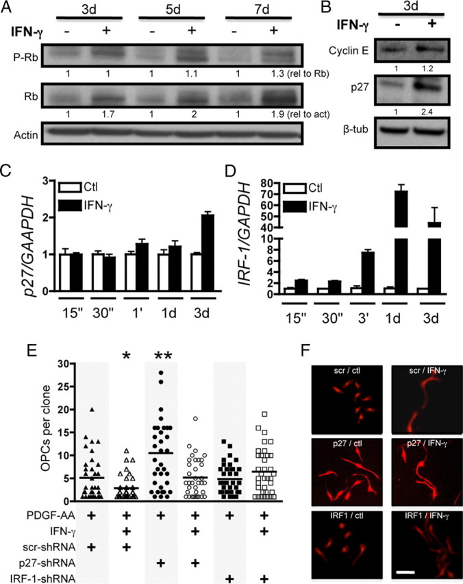
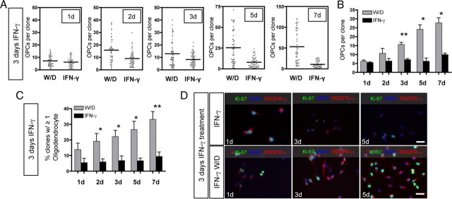
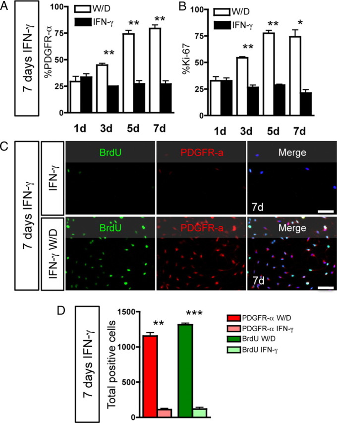
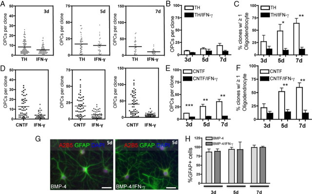
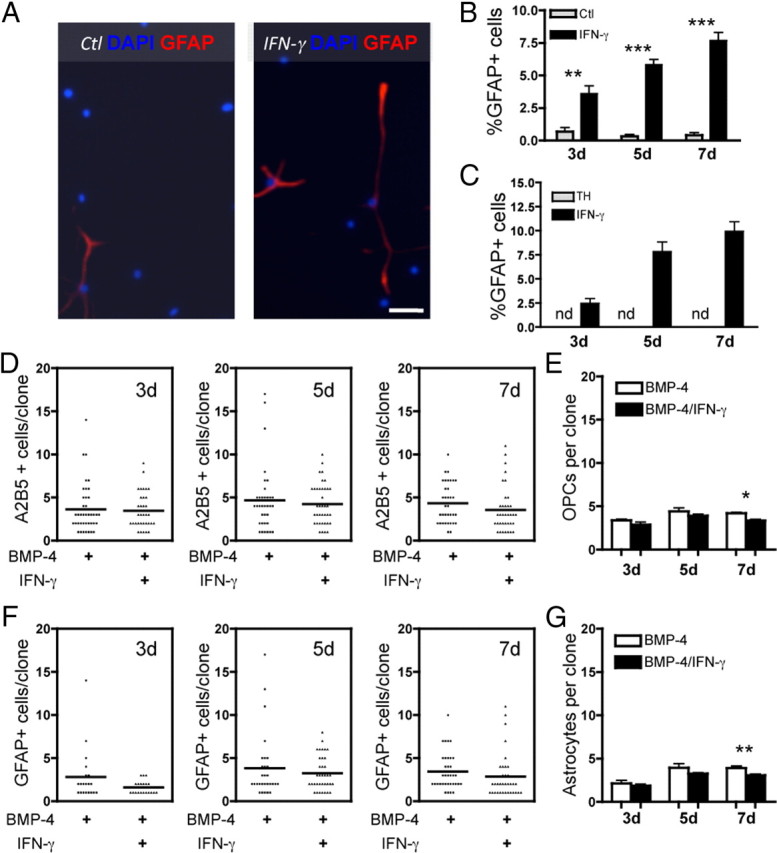
Oligodendrocyte-type 2 astrocyte progenitor cells (O-2A/OPCs) populate the CNS and generate oligodendrocytes and astrocytes in vitro and in vivo. Understanding how O-2A/OPCs respond to their environment is crucial to understanding how these cells function in the CNS and how to best promote their therapeutic proliferation and differentiation. We show that interferon-γ (IFN-γ) was not toxic to highly purified perinatal or adult rat O-2A/OPCs. IFN-γ treatment led to downregulation of PDGFR-α (platelet-derived growth factor receptor-α) and Ki-67 and decreased self-renewal in clonal populations. IFN-γ also significantly increased the proportion of cells in the G(0)/G(1) phase of the cell cycle, decreased BrdU (5-bromo-2'-deoxyuridine) incorporation, and led to increased expression of the cell cycle inhibitors Rb and p27(kip1). Although p27(kip1) expression was not necessary for IFN-γ-mediated quiescence, its upstream regulator IRF-1 was required. The quiescent state of O-2A/OPCs caused by IFN-γ was reversible as the withdrawal of IFN-γ allowed O-2A/OPCs to appropriately respond to both proliferation and differentiation signals. Differentiation into oligodendrocytes induced by either thyroid hormone or CNTF was also abrogated by IFN-γ. This inhibition was specific to the oligodendrocyte pathway, as O-2A/OPC differentiation into astrocytes was not inhibited. IFN-γ alone also led to the generation of GFAP-positive astrocytes in a subset of O-2A/OPCs. Together, these results demonstrate a reversible inhibitory effect of IFN-γ on O-2A/OPC proliferation with a concomitant generation of astrocytes. We propose that neuroinflammation involving increased IFN-γ can reduce progenitor numbers and inhibit differentiation, which has significant clinical relevance for injury repair, but may also contribute to the generation of astrocytes.


Figures

Figure 1.
Figure 1.
IFN-γ treatment decreases PDGFR-α and Ki-67 expression. A, O-2A/OPCs were grown at mass culture density in proliferation media (DMEM/F12 media with Sato components supplemented with 10 ng/ml PDGF-AA) with or without IFN-γ (10 ng/ml). O-2A/OPCs were stained for TUNEL. Cells were treated with increasing concentrations of IFN-γ (1, 5, 10, 20, 100 ng/ml), and as a control for cell death some cells were treated with 5 μm tert-butyl hydroperoxide. Error bars indicate mean ± SEM of two independent experiments. **p < 0.001, relative to control; ns, no significant difference; ANOVA followed by Bonferonni's post test. B, Whole-cell lysates were probed by Western blot for PDGFR-α and β-tubulin expression. C, D, Control-treated (C) and IFN-γ-treated (D) cells were immunostained for PDGFR-α and Ki-67. The open arrowhead represents a double-positive cell, the closed arrowhead represents a PDGFR-α-only positive cell, and the arrow represents a Ki-67-only positive cell. Scale bar, 25 μm. E, F, PDGFR-α-positive cells (E) and Ki-67-positive cells (F) were counted and reported as percentage positive of the total number of DAPI-positive cells. There were significantly fewer PDGFR-α- and Ki-67-positive cells after treatment with IFN-γ versus control at each time point. ***p < 0.001, **p < 0.01, t test.
Figure 2.
Figure 2.
Interferon-γ treatment decreases O-2A/OPC self-renewal. O-2A/OPCs were cultured at clonal density in basic growth media (DMEM/F12 with Sato components supplemented with 10 ng/ml of the mitogen PDGF-AA as described in Materials and Methods). bFGF (10 ng/ml) was also added as indicated. Cells were treated with or without IFN-γ (10 ng/ml) and stained with anti-A2B5, anti-GalC, and DAPI at the indicated time points, and clonal composition was determined. Cells were counted as O-2A/OPCs if they were A2B5+/GalC−. A–C, At 3 d (A), 5 d (B), and 7 d (C), O-2A/OPCs treated with PDGF-AA alone or in combination with bFGF gave rise to a significantly greater number of O-2A/OPCs per clone than those grown in the presence of IFN-γ (***p < 0.0001, ANOVA; *p < 0.05, ***p < 0.001, Bonferroni's post test). D, Quantification of A–C represented as mean ± SD and number of clones counted per treatment. A–D, Representative data of one experiment. E, Summary of data from four independent experiments. Means of individual experiments were grouped and data presented as mean ± SEM. IFN-γ exposure significantly reduced the number of O-2A/OPCs per clone at 3 d (**p < 0.001, ANOVA, followed by Bonferroni's post test), 5 d (***p < 0.001, ANOVA, followed by Bonferroni's post test), and 7 d (***p < 0.001, ANOVA, followed by Bonferroni's post test). F, O-2A/OPCs were grown at mass culture density and treated as indicated for 5 d and stained for PDGFR-α expression. Data are presented as mean ± SEM (***p < 0.0001, ANOVA, followed by Bonferroni's post test, **p < 0.001). G, O-2A/OPCs were grown at clonal density for 5 d as indicated and A2B5+ cells were counted per clone. Data are presented as mean ± SEM (***p < 0.0001, ANOVA, followed by Bonferroni's post test, **p < 0.001).
Figure 3.
Figure 3.
IFN-γ treatment leads to cell cycle exit. A, O-2A/OPCs were grown at mass culture density and treated with IFN-γ for 3 d. Cells were pulsed for 20 h with BrdU before staining. Scale bar, 25 μm. B, C, IFN-γ treatment significantly decreased BrdU incorporation (***p < 0.001, t test) (B) and PDGFR-α expression (***p < 0.001, t test) (C). D, IFN-γ significantly alters the proportion of O-2A/OPCs in each phase of the cell cycle as analyzed by flow cytometry. Cells were grouped by phase of the cell cycle and expressed as percentage of total cell number. The proportion of O-2A/OPCs in G0/G1 was significantly upregulated (**p < 0.005, t test), whereas the proportion in G2/M and S phase were both significantly decreased (*p < 0.01, t test). Representative examples of ModFit cell cycle analysis for control-treated cells (D′) and IFN-γ-treated cells (D″) with insets of representative scatterplots. E, F, IFN-γ decreases EdU incorporation and DNA content. O-2A/OPCs were analyzed by flow cytometry as above with a different DNA content dye and the thymidine analog EdU. Numbers represent the proportion of cells in each quadrant. G–I, Adult O-2A/OPCs were isolated from optic nerve as described and grown in culture for 4 d as indicated. G, Cells were pulsed with EdU for 48 h (days 3 and 4 in culture). Adult progenitors exposed to IFN-γ incorporated significantly less EdU than did controls (*p < 0.05, t test). H, Adult progenitors were plated at clonal density and maintained as described for 9 d in culture. Almost no clones exposed to IFN-γ had more than one cell at 9 d, whereas control-treated cells had significantly more clones with more than one cell (*p < 0.05, t test). I, Adult progenitors exposed to IFN-γ showed no difference in the proportion of GalC+ cells, although it was notable that all GalC+ cells were also A2B5+. Error bars represent mean ± SEM.
Figure 4.
Figure 4.
IRF-1 is required for IFN-γ-mediated quiescence. A, Whole-cell lysates obtained from O-2A/OPCs treated for indicated times were probed for phospho-Rb, quantified, and normalized against total Rb expression. Total Rb protein was quantified and normalized to actin intensity. B, Western blot analysis was performed for cyclin E and p27 and normalized to β-tubulin. C, D, O-2A/OPCs were treated for the indicated times with control or IFN-γ and RNA was isolated for RT-qPCR of p27, IRF-1, and GAPDH. Error bars represent mean ± SEM. p27 expression increased by 1 h but was only significant at 3 d (p < 0.001, t test vs control-treated cells). IRF-1 gene expression was significantly greater at each time point tested (p < 0.05, p < 0.001, t test vs control-treated cells). E, O-2A/OPCs grown at clonal density were infected with lentivirus-expressing shRNA constructs against p27, IRF-1, and a scrambled nontargeting control before control or IFN-γ treatment. IFN-γ significantly decreased self-renewal in scrambled shRNA infected cells (*p < 0.05, t test). p27 shRNA lentivirus infection had a significant effect on self-renewal in control-treated cells (**p < 0.001, t test vs control-treated scrambled shRNA-infected cells). IFN-γ treatment reduced self-renewal in p27 shRNA-treated cells to control levels. IRF-1 shRNA abrogated IFN-γ-mediated quiescence. F, Representative images of clones under each condition. Scale bar, 25 μm.
Figure 5.
Figure 5.
Withdrawal of IFN-γ enables O-2A/OPC self-renewal and spontaneous differentiation. O-2A/OPCs were grown at clonal density and treated with IFN-γ (10 ng/ml) and 10 ng/ml PDGF-AA for 3 d. W/D refers to cultures in which IFN-γ was withdrawn after 3 d and media was replaced with control proliferation media (DMEM/F12 media with Sato components and PDGF-AA; 10 ng/ml) for the given number of days indicated. IFN-γ treatment consisted of 3 d of IFN-γ followed by additional IFN-γ treatment for the number of days indicated. A, At 1 d after IFN-γ withdrawal, proliferation of O-2A/OPCs was not different from cells exposed to IFN-γ for 1 more day. At all other time points tested (as indicated), there was a significant increase in the number of OPCs per clone after IFN-γ withdrawal (2 d, p < 0.0001; 3 d, p = 0.0021; 5 d, p < 0.0001; 7 d, p < 0.0001, respectively, t test vs IFN-γ treatment). Data represent one experiment. B, Summary of data from multiple experiments. Means of individual experiments were grouped and data presented as mean ± SEM. IFN-γ withdrawal significantly increased the number of O-2A/OPCs per clone (*p < 0.05, **p < 0.01, t test). C, Summary data of multiple experiments in which clones from each time point were analyzed for GalC-expressing oligodendrocytes and are reported as the percentage of clones with at least one GalC-positive cell. There were significantly more clones with oligodendrocytes after IFN-γ withdrawal at each time point after 1 d (*p < 0.05, **p < 0.01, t test). D, Representative images of similar experiments grown at mass culture density for 3 d with IFN-γ followed by withdrawal of IFN-γ (bottom panel) and with IFN-γ (top panel) for another 1, 3, and 5 d. Cells were stained with anti-Ki-67, anti-PDGFR-α, and DAPI. Scale bar, 25 μm.
Figure 6.
Figure 6.
Long-term IFN-γ-induced quiescence is reversible. O-2A/OPCs were grown at mass culture density and treated with IFN-γ for 7 d. In separate cultures, IFN-γ was withdrawn and replaced with proliferation media (W/D) or growth in IFN-γ was continued for the same number of days (IFN-γ). A, At 3, 5, and 7 d W/D, there was significant upregulation of PDGFR-α expression (**p < 0.01, t test). B, Ki-67 expression was significantly upregulated in the W/D condition versus continuous IFN-γ at 3, 5, and 7 d (*p < 0.05, **p < 0.001, t test). C, BrdU incorporation and PDGFR-α expression were measured in O-2A/OPCs grown at mass culture density after W/D or continued IFN-γ. Scale bar, 50 μm. D, On W/D, both PDGFR-α expression and BrdU incorporation were significantly greater compared with continuous IFN-γ (**p < 0.005, ***p < 0.001, t test). Note that, in the 7 d IFN-γ group, these cells were exposed to IFN-γ for a total of 14 d and remained alive and quiescent. Error bars represent mean ± SEM.
Figure 7.
Figure 7.
O-2A/OPC differentiation into oligodendrocytes, but not astrocytes, is inhibited in the presence of IFN-γ. A, O-2A/OPCs were grown at clonal density in oligodendrocyte differentiation conditions (1 ng/ml PDGF-AA and 40 nm TH) in the presence or absence IFN-γ (10 ng/ml). There were significantly fewer O-2A/OPCs per clone found in differentiation conditions with the addition of IFN-γ at 3 d compared with control (***p < 0.001, t test), no difference at 5 d, and a significant decrease in O-2A/OPCs after 7 d of differentiation treatment with IFN-γ treatment compared with control (***p < 0.001, t test). Representative data from one experiment are shown. B, Means of individual experiments were grouped, and data are presented as mean ± SEM. IFN-γ exposure had no effect on O-2A/OPC proliferation in the presence of TH at any of the time points studied. C, Summary graph of multiple experiments in which GalC-positive oligodendrocytes were counted at each time point. At 3 d, there was no difference in the number of clones with at least one GalC-positive oligodendrocytes in the presence or absence of IFN-γ. By 5 d, there was a significant decrease in the number of clones containing oligodendrocytes (*p < 0.05, t test) and a greater decrease by 7 d (**p < 0.01, t test). D, O-2A/OPCs were induced to differentiate into oligodendrocytes with CNTF (10 ng/ml CNTF and 1 ng/ml PDGF-AA) and similar inhibition of differentiation was observed. The addition of IFN-γ to CNTF-treated O-2A/OPCs significantly decreased self-renewal at each time point (***p < 0.001, t test). Data are from one experiment. E, Means of individual experiments were grouped, and data are presented as mean ± SEM. There was a significant decrease in clones containing at least one GalC-positive oligodendrocyte in IFN-γ-treated cultures at 3, 5, and 7 d (**p < 0.01, ***p < 0.001, t test). F, Summary graph of multiple experiments in which GalC-positive oligodendrocytes were counted at each time point. There were significantly fewer clones containing oligodendrocytes at 5 and 7 d with IFN-γ treatment (**p < 0.01, t test). G, O-2A/OPCs were grown at mass culture density in the presence of the proastrocyte factor BMP-4 (10 ng/ml; and 1 ng/ml PDGF-AA) with or without IFN-γ (10 ng/ml). Type 2 astrocytes (A2B5 and GFAP immunoreactive) were generated at equal frequency in both conditions. H, Type 2 astrocytes were quantified from mass culture experiments as in E and are represented as the percentage GFAP-positive cells per total DAPI-positive nuclei. There is no difference in the proportion of type 2 astrocytes generated with or without IFN-γ. Scale bar, 25 μm.
Figure 8.
Figure 8.
IFN-γ promotes O-2A/OPC differentiation into GFAP-positive astrocytes. A, O-2A/OPCs were grown at mass culture density in minimal media (DMEM/F12 with Sato components supplemented with 1 ng/ml PDGF) and treated with or without IFN-γ (10 ng/ml). Cells were stained with anti-GFAP and DAPI at 5 d. Scale bar, 50 μm. B, Quantification of O-2A/OPC differentiation into GFAP-positive astrocytes at 3, 5, and 7 d. There was a significant increase in astrocyte differentiation in the presence of IFN-γ versus control (3 d, **p < 0.005; 5 and 7 d, ***p < 0.0001, t test). Note that the percentage of GFAP-positive cells in control was never more than 0.67%. C, O-2A/OPCs were grown at mass culture density in oligodendrocyte differentiation media (DMEM/F12 with Sato components supplemented with 1 ng/ml PDGF and 40 nm TH) and treated with or without IFN-γ (10 ng/ml). In the presence of thyroid hormone alone, no GFAP-positive cells were detected at any time point examined, but in the presence of IFN-γ, astrocyte generation is slightly higher than in B. D, O-2A/OPCs are responsible for generating IFN-γ-induced astrocytes. O-2A/OPCs were grown at clonal density in astrocyte differentiation media with or without IFN-γ (10 ng/ml). O-2A/OPC self-renewal, measured by the number of A2B5-positive/GalC-negative OPCs per clone, was not increased in by the addition of IFN-γ. Data are from one experiment. E, Summary graph of means from individual experiments grouped and data presented as mean ± SEM. IFN-γ did not increase self-renewal at any time point and actually led to fewer O-2A/OPCs per clone at 7 d (*p < 0.05, t test). F, Similar clonal cultures were stained with anti-GFAP, and the number of GFAP-positive cells per clone was quantified. There was no increase in proliferation of GFAP cells per clone with IFN-γ. Note the low numbers of cells per clone, indicating that there is no increase in astrocyte proliferation, although there is almost complete differentiation of all of the precursors into astrocytes. Data are from one experiment. G, Summary graph of means from individual experiments grouped and data presented as mean ± SEM. IFN-γ did not increase astrocyte proliferation at any time point and actually led to fewer astrocytes per clone at 7 d (**p < 0.001, t test).

Similar articles

Cited by

References

    1. Agresti C, D'Urso D, Levi G. Reversible inhibitory effects of interferon-gamma and tumour necrosis factor-alpha on oligodendroglial lineage cell proliferation and differentiation in vitro. Eur J Neurosci. 1996;8:1106–1116. - PubMed
    1. Aguirre A, Gallo V. Postnatal neurogenesis and gliogenesis in the olfactory bulb from NG2-expressing progenitors of the subventricular zone. J Neurosci. 2004;24:10530–10541. - PMC - PubMed
    1. Alonso G. NG2 proteoglycan-expressing cells of the adult rat brain: possible involvement in the formation of glial scar astrocytes following stab wound. Glia. 2005;49:318–338. - PubMed
    1. Andrews T, Zhang P, Bhat NR. TNFalpha potentiates IFNgamma-induced cell death in oligodendrocyte progenitors. J Neurosci Res. 1998;54:574–583. - PubMed
    1. Baerwald KD, Popko B. Developing and mature oligodendrocytes respond differently to the immune cytokine interferon-gamma. J Neurosci Res. 1998;52:230–239. - PubMed

Substances