. 2011 Jun 15;22(12):2119-30.
doi: 10.1091/mbc.E10-12-0969. Epub 2011 Apr 20.

GRB2 couples RhoU to epidermal growth factor receptor signaling and cell migration

Affiliations

GRB2 couples RhoU to epidermal growth factor receptor signaling and cell migration

Jin-San Zhang et al. Mol Biol Cell. .

Abstract

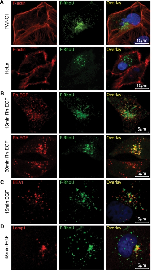
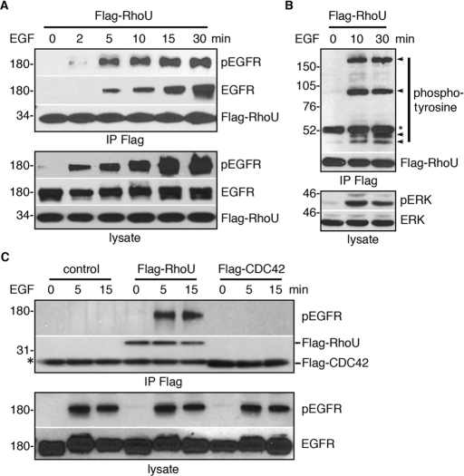
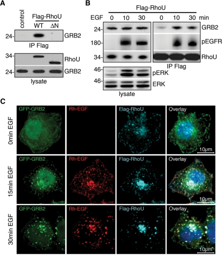
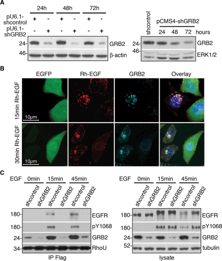
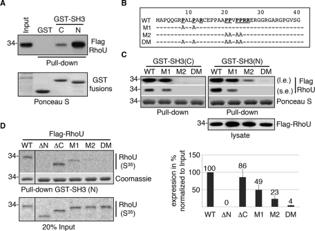
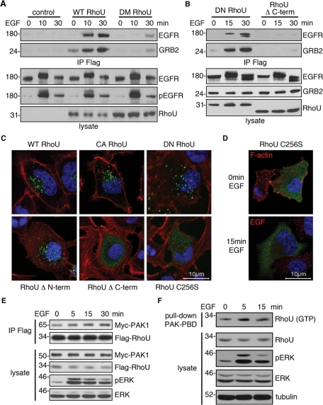
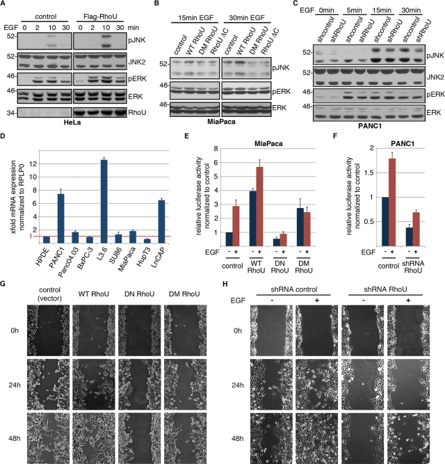
RhoU is an atypical Rho family member with high homology to CDC42 but containing unique N- and C-terminal extensions. The mechanisms regulating RhoU activation, as well as its downstream effectors, are not fully characterized. We show that after epidermal growth factor (EGF) stimulation RhoU colocalizes with EGF receptor (EGFR) on endosomes, which requires both its N- and C-terminal extension sequences. Moreover, RhoU physically associates with activated EGFR in a GRB2-dependent manner through specific proline-rich motifs within its N-terminus. Mutation of these proline-rich sequences or suppression of GRB2 by RNA interference abrogates the interaction of RhoU with activated EGFR, as well as EGF-stimulated RhoU GTP binding. In addition, RhoU is involved in EGFR-mediated signaling, leading to AP1 transcriptional activity and cell migration in pancreatic cancer cells, events that require its interaction with the Grb2-EGFR complex. Taken together, the data suggest a unique regulatory mechanism by which RhoU interaction with SH3 adaptor proteins might serve to integrate growth factor receptor signaling with RhoU activation.


Figures

FIGURE 1:
FIGURE 1:
RhoU localizes to endosomes and colocalizes with EGFR on EGF stimulation. (A) Immunofluorescence staining of PANC1 and HeLa cells transfected with a low level of Flag–RhoU were stained with anti-Flag (green), phalloidin (red) for F-actin, and Hoechst 33342 (blue) for DNA. (B) Flag-RhoU–transfected and serum-starved HeLa cells were stimulated with Rh-EGF (10 ng/ml; red) and stained with anti-Flag (green). (C, D) HeLa cells were stimulated with EGF (20 ng/ml) and stained with anti-Flag (green), anti-EEA1 (red, C), or anti-LAMP1 (red, D), and Hoechst 33342 (blue) for DNA.
FIGURE 2:
FIGURE 2:
RhoU, but not CDC42, physically associates with phosphorylated EGFR complex. (A, B) Flag-RhoU–transfected, serum-starved HeLa cells were stimulated with 20 ng/ml of EGF for the indicated time. Cells were lysed, immunoprecipitated, and immunoblotted with indicated antibodies. Arrows indicate tyrosine phosphorylated proteins coimmunoprecipitated with Flag–RhoU following EGF stimulation. Asterisk corresponds to nonspecific band from mouse immunoglobulin (Ig) G heavy chain. (C) HeLa cells were transfected with the empty vector, Flag–RhoU, and Flag–CDC42, respectively, immunoprecipitated, and immunoblotted with indicated antibodies. Asterisk denotes mouse IgG light chain.
FIGURE 3:
FIGURE 3:
RhoU N-terminus–mediated interaction with GRB2 is enhanced by EGF stimulation. (A) HeLa cells were transiently transfected with empty vector, Flag–RhoU, and its N-terminal deletion mutant (ΔN) expression plasmids. Lysates were immunoprecipitated and immunoblotted as indicated. (B) HeLa cells transfected with Flag–RhoU were serum starved before treatment with 20 ng/ml of EGF for the indicated time. Lysates were immunoprecipitated and immunoblotted with indicated antibodies. Efficient EGF activation is documented by ERK phosphorylation in total lysate. (C) Immunofluorescence staining of HeLa cells cotransfected with Flag–RhoU and GRB2–eGFP expression plasmids. Serum-starved cells were stimulated with Rh-EGF (10 ng/ml; red) for the indicated time and stained with anti-Flag (cyan) and Hoechst 33342 (blue) for DNA.
FIGURE 4:
FIGURE 4:
GRB2 is essential in coupling RhoU to EGFR complex. (A) Western blot analysis of HeLa cells transfected with pU6-shGRB2, pCMS4-shGRB2, or respective scrambled control vectors for time-dependent GRB2 suppression as indicated. (B) The pCMS4-shGRB2–transfected cells were seeded on cover slips and allowed to grow for 48 h, followed by 16 h of serum starvation before stimulation with Rh-EGF (10 ng/ml; red) for the indicated time. Cells were stained for GRB2 (light blue) and Hoechst 33342 (blue) for DNA. (C) Western blot analysis of HeLa cells transfected with pU6-shGRB2 or scrambled control vector for 24 h, then sequentially transfected with Flag–RhoU for another 24 h, followed by serum starvation for 16 h before EGF stimulation (20 ng/ml). Lysates were precipitated with anti-Flag agarose. Immunoprecipitates and lysates were immunoblotted with indicated antibodies. Numbers on the left correspond to protein molecular weight marker in kilodaltons.
FIGURE 5:
FIGURE 5:
Proline-rich motifs in the N-terminus of RhoU mediate its interaction with GRB2 and the EGFR. (A) Lysates from Flag-RhoU–transfected HeLa cells were used for GST pull-down assays with either the N- or C-terminal SH3 domain of GRB2 (top). The same blot stained with Ponceau S documents the fusion protein input (bottom). (B). The amino acid sequence of mouse RhoU N-terminal extension. The two class II SH3-binding consensuses (PXXPXR) are underlined in bold. The mutations by substitution of prolines with alanines in each mutant are specified. (C) Lysates from WT and mutant Flag-RhoU–transfected cells were used for GST pull-down assays with GRB2 N- and C-terminal SH3 fusion proteins, designated GST–SH3(N) and GST–SH3(C), respectively. Top two panels show Flag–RhoU bound to GST–SH3 fusion proteins as detected by anti-Flag antibodies (l.e. and s.e. indicate longer and shorter exposure, respectively). Ponceau S staining documents the input of recombinant GST fusion proteins. The expression level of Flag–RhoU constructs is shown in the bottom panel. (D) GST SH3(N) was used in pull-down assay with in vitro–translated [35S]methionine-labeled RhoU containing indicated deletion/mutations. A representative result of RhoU proteins bound to GST SH3(N) is shown in the top panel followed by the Coomassie staining of the same gel to the indicated input of GST–SH3 fusion proteins. The input (20%) of in vitro–translated RhoU proteins is shown at the bottom. The average band intensity for GST-SH3–precipitated RhoU from three independent experiments along with SD is shown in the histogram (right).
FIGURE 6:
FIGURE 6:
The N- and C-terminal extensions, but not GTP loading, are required for RhoU–GRB2/EGFR interactions. (A, B) Western blot analysis of HeLa transfected with indicated Flag–RhoU expression vectors for 24 h, followed by serum starvation for 16 h before EGF stimulation (20 ng/ml). Equal amount of lysate was precipitated with anti-Flag agarose. Immunoprecipitates and lysates were immunoblotted with indicated antibodies. (C, D) Immunofluorescence staining of HeLa cells expressing the indicated RhoU constructs (green). Transfected cells were maintained in normal medium for 24 h (C) or serum starved for additional 16 h and stimulated with EGF stimulation (20 ng/ml) (D) before fixing and staining with anti-Flag (green), phalloidin (red) for F-actin, and Hoechst 33342 (blue) for DNA. (E) HeLa cells were cotransfected with Flag–RhoU and Myc–PAK1 for 24 h, followed by 16 h of serum starvation. Lysates were prepared after stimulation with EGF (50 ng/ml) for the indicated time, immunoprecipitated, and blotted with RhoU, myc, and additional antibodies as indicated. (F) HeLa cells were transfected with Flag–RhoU and treated with EGF as in E. Cell lysates were prepared for pull-down assay with GST–PBD. The pull-down samples and lysate were immunoblotted with indicated antibodies.
FIGURE 7:
FIGURE 7:
RhoU synergize with EGF in JNK/AP1 activation and cell migration. (A) HeLa cells transfected with control vector or Flag–RhoU were serum starved for 16 h before EGF stimulation (20 ng/ml) for the indicated period of time. Cell lysates were prepared and immunoblotted for activating phosphorylation of the major EGF-dependent signaling pathways with indicated antibodies. (B) Miapaca2 cells transfected with WT or mutant RhoU expression constructs were serum starved, treated with EGF (100 ng/ml). and immunoblotted with the indicated antibodies. (C) PANC1 cells with stable RhoU suppression or scramble control were analyzed for JNK phosphorylation following EGF treatment (100 ng/ml). Phosphorylated ERK documents successful EGF stimulation, and ERK staining reflects relatively even sample loading in A–C. (D) Quantitative RT-PCR for RhoU mRNA expression in immortalized human ductal epithelial cells (HPDEs), different pancreatic cancer cell lines, and the prostate cancer cell line LNCaP cells. Results are normalized to RPLP0 and HPDE cells. (E, F) Luciferase reporter assay for AP1 activity in low-level (Miapaca2, E) and high-level (PANC1, F) RhoU-expressing cells. Miapaca2 cells were transfected with the 6xAP1 luciferase reporter and indicated constructs for 24 h and subsequently treated with EGF (20 ng/ml) or DMSO for 24 h. PANC1 cells were transfected with shRNA against RhoU or control shRNA for 36 h and subsequently treated with EGF (20 ng/ml) or DMSO for 24 h. Results are mean of three independent experiments ±SD. (G) For wound-healing analysis PANC1 cells were transfected with indicated constructs for 12 h and serum starved for another 12 h. At a transfection efficiency of >70% a cell-free space was made by scraping through the monolayer using a pipette tip. (H) PANC1 cells stably expressing RhoU shRNA or scrambled control were seeded at a high density and serum starved overnight. Scratches were made as in G, and EGF 20 ng/ml or DMSO was supplemented as indicated. Wound closure was documented every 12 h. Results from one representative experiment of three independent studies are shown.

References

    1. Alan JK, Berzat AC, Dewar BJ, Graves LM, Cox AD. Regulation of the Rho family small GTPase Wrch-1/RhoU by C-terminal tyrosine phosphorylation requires Src. Mol Cell Biol. 2010;30:4324–4338. - PMC - PubMed
    1. Aspenström P, Fransson A, Saras J. Rho GTPases have diverse effects on the organization of the actin filament system. Biochem J. 2004;377:327–337. - PMC - PubMed
    1. Berzat AC, Buss JE, Chenette EJ, Weinbaum CA, Shutes A, Der CJ, Minden A, Cox AD. Transforming activity of the Rho family GTPase, Wrch-1, a Wnt-regulated Cdc42 homolog, is dependent on a novel carboxyl-terminal palmitoylation motif. J Biol Chem. 2005;280:33055–33065. - PubMed
    1. Brady DC, Alan JK, Madigan JP, Fanning AS, Cox AD. The transforming Rho family GTPase Wrch-1 disrupts epithelial cell tight junctions and epithelial morphogenesis. Mol Cell Biol. 2009;29:1035–1049. - PMC - PubMed
    1. Brazier H, Pawlak G, Vives V, Blangy A. The Rho GTPase Wrch1 regulates osteoclast precursor adhesion and migration. Int J Biochem Cell Biol. 2009;41:1391–1401. - PubMed

Publication types

MeSH terms