. 2011 Jun 15;22(12):1971-84.
doi: 10.1091/mbc.E11-01-0037. Epub 2011 Apr 20.

A yeast model for polyalanine-expansion aggregation and toxicity

Affiliations

A yeast model for polyalanine-expansion aggregation and toxicity

Catherine A Konopka et al. Mol Biol Cell. .

Abstract

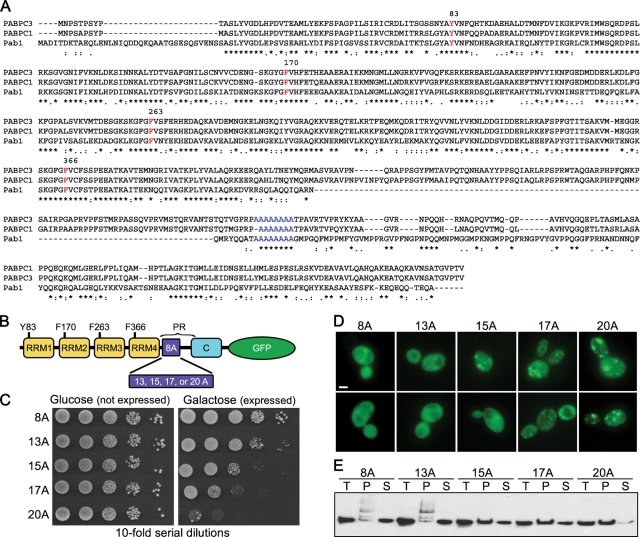
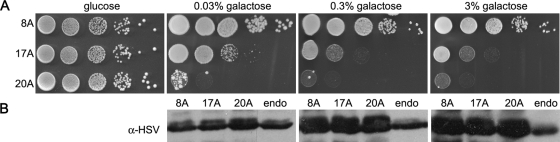
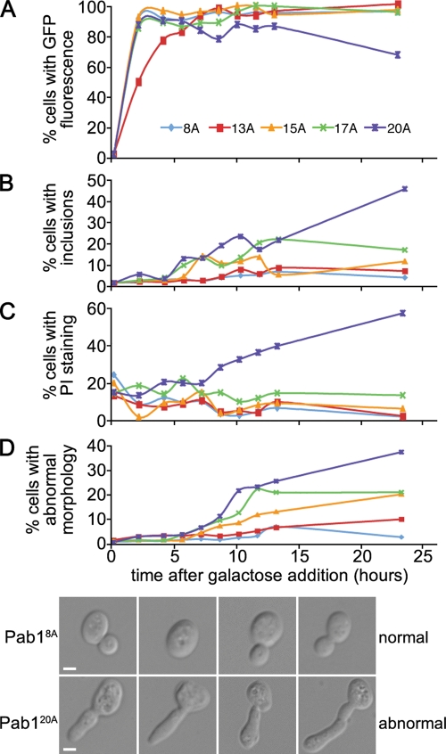
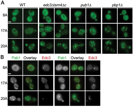
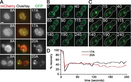
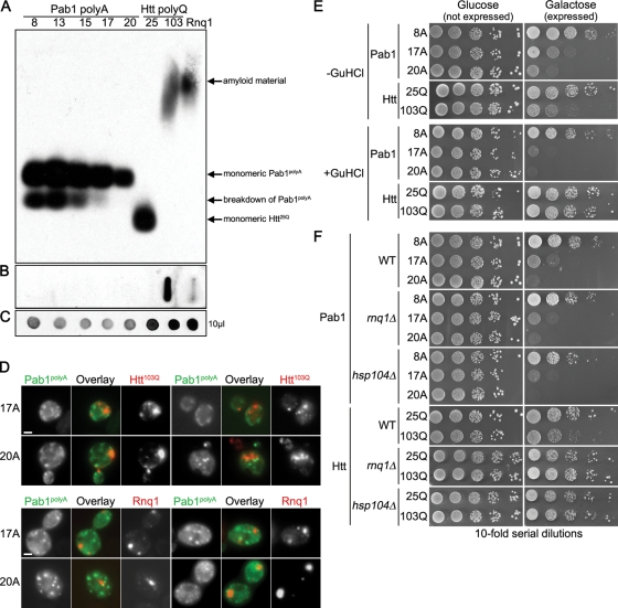
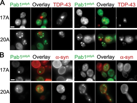
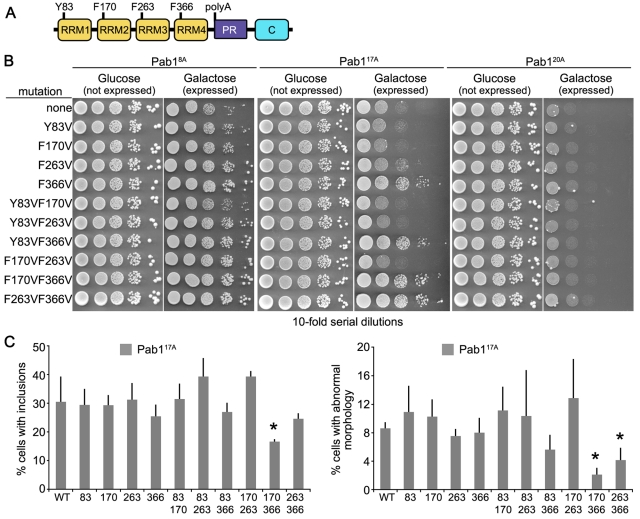
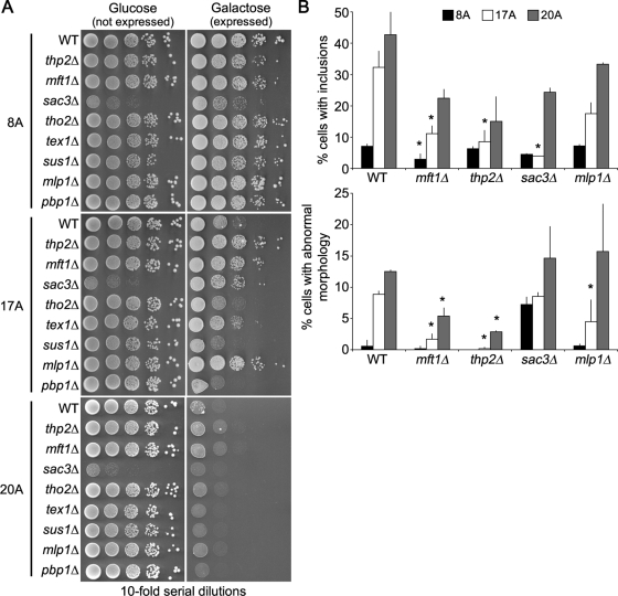
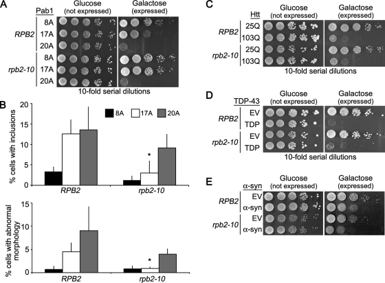
Nine human disorders result from the toxic accumulation and aggregation of proteins with expansions in their endogenous polyalanine (polyA) tracts. Given the prevalence of polyA tracts in eukaryotic proteomes, we wanted to understand the generality of polyA-expansion cytotoxicity by using yeast as a model organism. In our initial case, we expanded the polyA tract within the native yeast poly(Adenine)-binding protein Pab1 from 8A to 13A, 15A, 17A, and 20A. These expansions resulted in increasing formation of Pab1 inclusions, insolubility, and cytotoxicity that correlated with the length of the polyA expansion. Pab1 binds mRNA as part of its normal function, and disrupting RNA binding or altering cytoplasmic mRNA levels suppressed the cytotoxicity of 17A-expanded Pab1, indicating a requisite role for mRNA in Pab1 polyA-expansion toxicity. Surprisingly, neither manipulation suppressed the cytotoxicity of 20A-expanded Pab1. Thus longer expansions may have a different mechanism for toxicity. We think that this difference underscores the potential need to examine the cytotoxic mechanisms of both long and short expansions in models of expansion disorders.


Figures

FIGURE 1:
FIGURE 1:
Pab1 polyA-expansions are toxic and insoluble and aggregate in a polyA tract length–dependent manner. (A) Amino acid alignment of human PABPC1, PABPC3, and yeast Pab1. Residues required for RNA binding are indicated in red and the polyA tract in blue. (B) Schematic of Pab1 domain structure. C, C-terminal domain; PR, proline rich. (C) Tenfold serial dilutions of cells were spotted onto medium containing glucose (Pab1 expression repressed) or galactose (Pab1 expression induced) to measure spotting efficiency and toxicity of Pab1 polyA-expansions. (D) Pab1 expression (with indicated polyA tract length) was induced in wild-type cells for 16 h and imaged with epifluorescence microscopy for Pab1-GFP localization. Bar = 2 μm. (E) Solubility of Pab1 at 13,000 rcf. T, total; S, supernatant; P, pellet.
FIGURE 2:
FIGURE 2:
Expression levels of endogenous and PolyA-expanded Pab1. (A) Tenfold serial dilutions of cells were spotted onto medium containing glucose (polyA-expanded Pab1 expression repressed) or varying concentrations of galactose (polyA-expanded Pab1 expression induced) to measure spotting efficiency and toxicity of Pab1 polyA-expansions at increasing Pab1 expression levels. (B) Immunoblot with anti-HSV to detect Pab1 tagged at its native locus (endo) and Pab1 polyA-expansion variants grown in increasing galactose concentrations. Equal OD equivalents were loaded on the polyacrylamide gel.
FIGURE 3:
FIGURE 3:
Time course of polyA-expanded Pab1 expression, toxicity, and aggregation. (A–D) Time course of Pab1–GFP expression (A), aggregate formation (B), cell death (C), and morphology defects (D). Cell death was measured by the accumulation of propidium iodide (PI) in cells. Reported as a percentage of total cells (A, C, and D) or as a percentage of GFP-positive cells (B). (D) Brightfield images of cells expressing Pab18A-GFP or Pab120A-GFP, as indicated. Bar = 2 μm.
FIGURE 4:
FIGURE 4:
Pab1 polyA-expansion inclusions are not P-bodies or stress granules. (A) Pab18A-GFP, Pab117A-GFP, or Pab120A-GFP expression was induced for 16 h in the indicated strains defective in P-body (edc3Δlsm4Δc) or stress granule formation (pub1Δ and pbp1Δ) and imaged with epifluorescence microscopy. (B) As in (A), except Pab18A-GFP, Pab117A-GFP, or Pab120A-GFP was coexpressed with Edc3-mCherry in wild-type cells. Bars = 2 μm.
FIGURE 5:
FIGURE 5:
Pab117A and Pab120A inclusions do not contain Pab18A and do not exchange readily with the soluble pool. (A) Pab117A-GFP or Pab120A-GFP was coexpressed with Pab18A-mCherry and imaged with epifluorescence microscopy. (B and C) Images of FRAP time series, Pab117A-GFP (B) and Pab120A-GFP (C). Numbers represent time in seconds after start of imaging and correspond to graph in D. (D) Graph of FRAP experiments shown in B and C of Pab117A-GFP and Pab120A-GFP induced for 16 h. Bars = 2 μm.
FIGURE 6:
FIGURE 6:
Pab1 polyA-expansion aggregates are distinct from polyQ-expansion and prion aggregates. (A–C) Immunoblotting with anti-GFP to detect Pab1-GFP polyA-expansion variants, Htt-GFP polyQ-expansion variants, or Rnq1-YFP. (A) Semidenaturing detergent agarose gel electrophoresis comparing SDS solubility of Pab1 polyA-expansions with Htt polyQ-expansions and Rnq1. (B) Filter retardation assay showing the ability of Pab1 polyA-expansion variants and Htt25Q but not Htt103Q to pass through 0.2-μm cellulose acetate filter. (C) A total of 10 μl of total protein extract was spotted onto nitrocellulose to determine relative protein concentration. (D) Cells coexpressing Pab117A-GFP or Pab120A-GFP with either Htt103Q-mRFP or Rnq1-mCherry were imaged by epifluorescence microscopy. Bars = 2 μm. (E) Tenfold serial dilutions of cells were spotted onto medium containing glucose (expression repressed) or galactose (expression induced) to measure spotting efficiency and toxicity of polyA-expansion Pab1 or polyQ-expansion Htt after three successive passages on guanidine-HCl (GuHCl) to cure yeast of prions. (F) Tenfold serial dilutions of cells were spotted onto medium containing glucose (expression repressed) or galactose (expression induced) to measure spotting efficiency and toxicity of polyA-expansion Pab1 or polyQ-expansion Htt in hsp104Δ or rnq1Δ cells.
FIGURE 7:
FIGURE 7:
PolyA-expanded Pab1 inclusions are distinct from TDP-43 and α-syn inclusions. Cells coexpressing Pab117A-GFP or Pab120A-GFP with either TDP43-DsRed (A) or α-syn-mCherry (B) were imaged by epifluorescence microscopy. Bars = 2 μm.
FIGURE 8:
FIGURE 8:
Pab117A but not Pab120A requires RNA binding for toxicity and aggregation. (A) Schematic of Pab1 domain structure. C, C-terminal domain; PR, proline rich. (B) Tenfold serial dilutions of cells were spotted onto medium containing glucose (expression repressed) or galactose (expression induced) to measure spotting efficiency and toxicity of Pab117A and Pab120A RNA-binding mutants. (C) Cells expressing RNA-binding mutants of Pab117A were assayed for presence of GFP-positive inclusions and morphology defects after 16-h induction and reported as a percentage of GFP-positive cells. More than 200 cells in three independent cultures were counted for each. Bars are SD. *p < 0.05 by Student's t test when compared with wild-type Pab117A.
FIGURE 9:
FIGURE 9:
Pab117A but not Pab120A toxicity is suppressed in mRNA export mutants. (A) Tenfold serial dilutions of cells were spotted onto medium containing glucose (expression repressed) r galactose (expression induced) to measure spotting efficiency toxicity of Pab18A-GFP, Pab117A-GFP, or Pab120A-GFP in wild-type and deletion mutant cells. (B) Wild-type and deletion mutant cells expressing Pab18A-GFP, Pab117A-GFP, or Pab120A-GFP were assayed for presence of GFP-positive inclusions and morphology defects after 16-h induction and reported as a percentage of GFP-positive cells. More than 150 cells in three independent cultures were counted for each. Bars are SD. *p < 0.05 by Student's t test when polyA-expansion Pab1 mutant strains were compared with corresponding wild type.
FIGURE 10:
FIGURE 10:
Htt103Q and α-syn toxicity is not suppressed in mRNA export mutants, whereas TDP-43 toxicity is suppressed. Tenfold serial dilutions of cells were spotted onto medium containing glucose (expression repressed) or galactose (expression induced) to measure spotting efficiency and toxicity of Htt25Q and Htt103Q (A), TDP-43 or α-syn (B) in wild-type and deletion mutant cells.
FIGURE 11:
FIGURE 11:
Pab117A but not Pab120A toxicity is suppressed by globally lowering mRNA levels. (A) Tenfold serial dilutions of cells were spotted onto medium containing glucose (expression repressed) or galactose (expression induced) to measure spotting efficiency and toxicity of Pab117A-GFP or Pab120A-GFP in wild-type and RNA PolII mutant (rpb2–10) cells. (B) Wild-type and rpb2–10 cells expressing Pab18A-GFP, Pab117A-GFP, or Pab120A-GFP were assayed for presence of GFP-positive inclusions and morphology defects after 16-h induction and reported as a percentage of GFP-positive cells. More than 150 cells in three independent cultures were counted for each. Bars are SD. *p < 0.05 by Student's t test when Pab1polyA rpb2–10 strains were compared with corresponding RPB2 strain. (C–E) As in (A) except that cells expressing Htt25Q or Htt103Q (C), TDP-43-GFP (D), or α-syn (E) were assayed.

Similar articles

Cited by

References

    1. Abu-Baker A, Messaed C, Laganiere J, Gaspar C, Brais B, Rouleau GA. Involvement of the ubiquitin-proteasome pathway and molecular chaperones in oculopharyngeal muscular dystrophy. Hum Mol Genet. 2003;12:2609–2623. - PubMed
    1. Albrecht A, Mundlos S. The other trinucleotide repeat: polyalanine expansion disorders. Curr Opin Genet Dev. 2005;15:285–293. - PubMed
    1. Albrecht AN, Kornak U, Boddrich A, Suring K, Robinson PN, Stiege AC, Lurz R, Stricker S, Wanker EE, Mundlos S. A molecular pathogenesis for transcription factor associated poly-alanine tract expansions. Hum Mol Genet. 2004;13:2351–2359. - PubMed
    1. Amiel J, Trochet D, Clement-Ziza M, Munnich A, Lyonnet S. Polyalanine expansions in human. Hum Mol Genet. 2004;13 Spec No 2:R235–R243. - PubMed
    1. Amrani N, Minet M, Le Gouar M, Lacroute F, Wyers F. Yeast Pab1 interacts with Rna15 and participates in the control of the poly(A) tail length in vitro. Mol Cell Biol. 1997;17:3694–3701. - PMC - PubMed

Publication types

MeSH terms

LinkOut - more resources