Review
. 2011:30:22-60.
doi: 10.1159/000324065. Epub 2011 Apr 19.

Addiction and brain reward and antireward pathways

Affiliations
Review

Addiction and brain reward and antireward pathways

Eliot L Gardner. Adv Psychosom Med. 2011.

Abstract

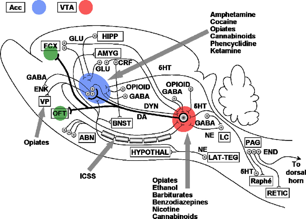
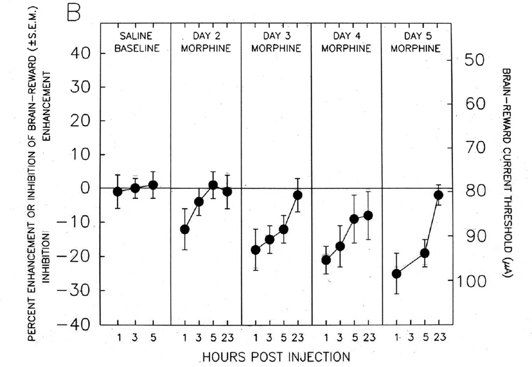
Addictive drugs have in common that they are voluntarily self-administered by laboratory animals (usually avidly), and that they enhance the functioning of the reward circuitry of the brain (producing the 'high' that the drug user seeks). The core reward circuitry consists of an 'in-series' circuit linking the ventral tegmental area, nucleus accumbens and ventral pallidum via the medial forebrain bundle. Although originally believed to simply encode the set point of hedonic tone, these circuits are now believed to be functionally far more complex, also encoding attention, expectancy of reward, disconfirmation of reward expectancy, and incentive motivation. 'Hedonic dysregulation' within these circuits may lead to addiction. The 'second-stage' dopaminergic component in this reward circuitry is the crucial addictive-drug-sensitive component. All addictive drugs have in common that they enhance (directly or indirectly or even transsynaptically) dop-aminergic reward synaptic function in the nucleus accumbens. Drug self-administration is regulated by nucleus accumbens dopamine levels, and is done to keep nucleus accumbens dopamine within a specific elevated range (to maintain a desired hedonic level). For some classes of addictive drugs (e.g. opiates), tolerance to the euphoric effects develops with chronic use. Postuse dysphoria then comes to dominate reward circuit hedonic tone, and addicts no longer use drugs to get high, but simply to get back to normal ('get straight'). The brain circuits mediating the pleasurable effects of addictive drugs are anatomically, neurophysiologically and neurochemically different from those mediating physical dependence, and from those mediating craving and relapse. There are important genetic variations in vulnerability to drug addiction, yet environmental factors such as stress and social defeat also alter brain-reward mechanisms in such a manner as to impart vulnerability to addiction. In short, the 'bio-psycho-social' model of etiology holds very well for addiction. Addiction appears to correlate with a hypodopaminergic dysfunctional state within the reward circuitry of the brain. Neuroimaging studies in humans add credence to this hypothesis. Credible evidence also implicates serotonergic, opioid, endocannabinoid, GABAergic and glutamatergic mechanisms in addiction. Critically, drug addiction progresses from occasional recreational use to impulsive use to habitual compulsive use. This correlates with a progression from reward-driven to habit-driven drug-seeking behavior. This behavioral progression correlates with a neuroanatomical progression from ventral striatal (nucleus accumbens) to dorsal striatal control over drug-seeking behavior. The three classical sets of craving and relapse triggers are (a) reexposure to addictive drugs, (b) stress, and (c) reexposure to environmental cues (people, places, things) previously associated with drug-taking behavior. Drug-triggered relapse involves the nucleus accumbens and the neurotransmitter dopamine. Stress-triggered relapse involves (a) the central nucleus of the amygdala, the bed nucleus of the stria terminalis, and the neurotransmitter corticotrophin-releasing factor, and (b) the lateral tegmental noradrenergic nuclei of the brain stem and the neurotransmitter norepinephrine. Cue-triggered relapse involves the basolateral nucleus of the amygdala, the hippocampus and the neurotransmitter glutamate. Knowledge of the neuroanatomy, neurophysiology, neurochemistry and neuropharmacology of addictive drug action in the brain is currently producing a variety of strategies for pharmacotherapeutic treatment of drug addiction, some of which appear promising.


Figures

Fig. 1
Fig. 1. BRAIN REWARD CIRCUITRY
Diagram of the brain-reward circuitry of the mammalian (laboratory rat) brain, with sites at which various addictive drugs act to enhance brain reward mechanisms and thus to produce drug-seeking behavior, drug-taking behavior, drug-craving, and relapse to drug-seeking behavior. ABN, anterior bed nuclei of the medial forebrain bundle; Acc, nucleus accumbens; AMYG, amygdala; DA, subcomponent of the ascending mesocorticolimbic dopaminergic system that appears to be preferentially activated by addictive drugs; DYN, dynorphinergic neuronal fiber bundle outflow from the nucleus accumbens; ENK, enkephalinergic neuronal fiber bundle outflow from the nucleus accumbens; FCX, frontal cortex; GABA, GABAergic inhibitory fiber systems synapsing in the ventral tegmental area, the nucleus accumbens, and into the vicinity of the locus coeruleus, as well as the GABAergic neuronal fiber bundle outflow from the nucleus accumbens; GLU, glutamatergic neural systems originating in frontal cortex and synapsing in both the ventral tegmental area and the nucleus accumbens; 5HT, serotonergic (5-hydroxytryptamine) fibers, which originate in the anterior raphé nuclei and project to both the cell body region (ventral tegmental area) and terminal projection field (nucleus accumbens) of the DA reward neurons; ICSS, the descending, myelinated, moderately fast conducting component of the brain-reward circuitry that is preferentially activated by electrical intracranial self-stimulation (electrical brain-stimulation reward); LC, locus coeruleus; NE, noradrenergic fibers, which originate in the locus coeruleus and synapse into the general vicinity of the ventral mesencephalic neuronal cell fields of the ventral tegmental area; Opioid, endogenous opioid peptide neural systems synapsing into both the ventral tegmental DA cell fields and the nucleus accumbens DA terminal projection loci; Raphé, brainstem serotonergic raphé nuclei; VP, ventral pallidum; VTA, ventral tegmental area. After [1].
Fig. 2
Fig. 2. LEFT SHIFT WITH ADDICTIVE DRUG, COUNTERED BY SELECTIVE D3 RECEPTOR ANTAGONIST
Enhancement of electrical brain-stimulation reward by an addictive drug (nicotine) and attenuation of that enhancement by a highly selective dopamine D3 receptor antagonist (SB-277011A). Left-shifts in such rate-frequency brain-reward functions denote enhancement of brain reward (the drug-induced “high”); right-shifts denote inhibition of brain reward. After [38].
Fig. 3
Fig. 3. REAL-TIME DOPAMINE BRAIN MICRODIALYSIS DURING INTRAVENOUS OPIATE SELF-ADMINISTRATION
Minute-by-minute measurements - using in vivo brain microdialysis - of extracellular nucleus accumbens dopamine in a laboratory rat voluntarily self-administering intravenous heroin. The thick dashed vertical lines indicate the start (left thick dashed vertical line) and end (right thick dashed vertical line) of availability of heroin. The thin solid vertical lines indicate a voluntary self-administration of heroin (a heroin “hit”) by the test animal. At the beginning of every drug self-administration session, extracellular nucleus accumbens dopamine is tonically enhanced approximately 200% by the initial “hit(s)” of heroin. Thereafter, extracellular nucleus accumbens dopamine fluctuates phasically - with the animal becoming motivated to give itself another drug “hit” whenever extracellular nucleus accumbens dopamine drops to approximately 100% over its initial pre-heroin level. Thus, the animal appears to self-administer the addictive drug to enhance extracellular nucleus accumbens dopamine and to keep it within a preferred (and elevated) range – thus keeping hedonic tone enhanced over its normal basal state when unaffected by addictive drugs. After [49,50].
Fig. 4
Fig. 4. PRO-REWARD AND ANTI-REWARD BRAIN-STIMULATION REWARD SUBSTRATES ACTIVATED BY OPIATE ADMINISTRATION - FROM NAZZARO AND GARDNER
Brain reward and anti-reward mechanisms recruited by systemic opiate administration. Enhancement or inhibition of brain-reward is measured using electrical brain-stimulation reward in laboratory rats. The stimulating electrode in Panel A is located in the medial portion of the mesotelencephalic dopaminergic reward fiber tract; the stimulating electrode in Panel B is located in the lateral portion of the mesotelencephalic dopaminergic reward fiber tract. Panel A: Acute systemic opiate administration enhances brain reward, which diminishes as opiate effects wear off during each test day, and which shows development of tolerance with successive opiate administrations. Panel B: Acute systemic opiate administration inhibits brain reward, which diminishes as opiate effects wear off during each test day, and which shows development of sensitization (enhancement, “reverse tolerance”) with successive opiate administrations. Overall hedonic tone is a combination of effects depicted in Panels A and B. Thus, with chronic opiate administration, the reward-enhancing effects diminish and the anti-reward effects intensify, producing and overall reward deficiency state. After [42,58].
Fig. 5
Fig. 5. RANK IN A SOCIAL HIERARCHY CONTRIBUTES TO BRAIN DA DEFICIENCY AND VULNERABILITY TO ADDICTIVE DRUG-TAKING BEHAVIOR
Intravenous cocaine self-administration in monkeys of differing social hierarchy status within the social group. The solid black circles indicate monkeys with low social status; the open circles indicate moneys with high social status. Low social status produces a dopamine deficit in the mesotelencephalic reward circuitry, and a proclivity to self-administer addictive drugs. After [77].
Fig. 6
Fig. 6. TIME-LINE GRAPH OF REINSTATEMENT (RELAPSE) TO ADDICTIVE DRUG-SEEKING BEHAVIOR
Schematic diagram of the “reinstatement” animal model of relapse to drug-seeking behavior. Initially, animals are allowed to freely self-administer intravenous cocaine in the presence of environmental cues that indicate the availability of cocaine. At the point indicated by the first vertical dashed line, the cue lights are turned off and saline is substituted for cocaine in the infusion apparatus. This causes behavioral extinction of the drug-taking habit and, perforce, pharmacological “detoxification” from cocaine. On the last day of the extinction phase of the experiment, the animal is given a single non-contingent intravenous “hit” of cocaine. This causes immediate and robust relapse to intense levels of drug-seeking behavior (although the infusion apparatus is filled with saline, as during the extinction and detoxification stage). After [37].
Fig. 7
Fig. 7. INCUBATION OF DRUG CRAVING
Incubation (enhancement) of drug-craving with the mere passage of time. Panel A: Drug-seeking behavior measured as extinction responding. Panel B: Drug-seeking behavior measured as cue-triggered relapse to drug seeking behavior. After [169].

References

    1. Gardner EL. Brain reward mechanisms. In: Lowinson JH, Ruiz P, Millman RB, Langrod JG, editors. Substance Abuse: A Comprehensive Textbook. 4th edn. Philadelphia: Lippincott Williams & Wilkins; 2005. pp. 48–97.
    1. Gardner EL. What we have learned about addiction from animal models of drug self-administration. Am J Addict. 2000;9:285–313. - PubMed
    1. O’Brien CP, Gardner EL. Critical assessment of how to study addiction and its treatment: human and non-human animal models. Pharmacol Ther. 2005;108:18–58. - PubMed
    1. Gardner EL, David J. The neurobiology of chemical addiction. In: Elster J, Skog O-J, editors. Getting Hooked: Rationality and the Addictions. Cambridge, England: Cambridge University Press; 1999. pp. 93–136.
    1. Wise RA, Gardner EL. Functional anatomy of substance-related disorders. In: D'haenen H, den Boer JA, Willner P, editors. Biological Psychiatry. New York: Wiley; 2002. pp. 509–522.

MeSH terms

Substances