

Targeting lymphangiogenesis after islet transplantation prolongs islet allograft survival
- PMID: 21508896
- PMCID: PMC3703312
- DOI: 10.1097/TP.0b013e31821d2661
Targeting lymphangiogenesis after islet transplantation prolongs islet allograft survival
Abstract
Background: Lymphatics are important for their conduit functions of transporting antigen, immune cells, and inflammatory mediators to draining lymph nodes and to the general circulation. Lymphangiogenesis is involved in many pathologic processes; however, the roles for lymphatic responses in transplantation have not been thoroughly investigated.
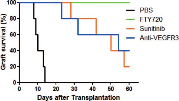
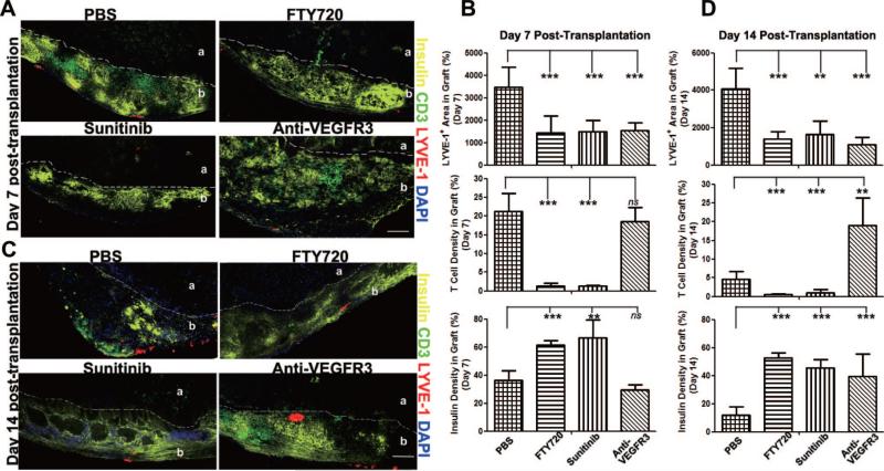
Methods: Mice were made diabetic by a single high dose of streptozotocin and then received islet allografts. Animals were treated with three different lymphatic inhibitors. FTY720, an analog of sphingosine 1-phosphate, inhibited lymphocyte migration into afferent and efferent lymphatics. Sunitinib, a kinase inhibitor, blocked several receptors, including vascular endothelial growth factor receptor 3 (VEGFR3), the major growth factor receptor for lymphatic endothelial cells. Anti-VEGFR3 monoclonal antibody specifically inhibited VEGFR3. Diabetes was determined by daily monitoring of blood glucose levels. Inflammation within islet grafts was assessed by immunohistochemistry for insulin, T cells (CD3), and lymphatics (LYVE-1).
Results: After transplantation, lymphangiogenesis occurred in islet allografts and in draining lymph nodes. FTY720, sunitinib, and anti-VEGFR3 each inhibited lymphangiogenesis in the islets and significantly prolonged allograft survival. Immunofluorescent staining demonstrated that administration of each of the lymphatic inhibitors resulted in preservation of islets and β-cells along with a markedly reduced infiltration of T cells into the grafts.
Conclusion: Lymphangiogenesis occurs in islet allografts in response to inflammation and plays a key role in the islet inflammation in alloimmunity. Interfering with lymphatic function leads to inhibition of lymphangiogenesis and prolonged or indefinite allograft survival. These observations suggest new therapeutic targets for rejection and tolerance.
Figures


References
-
- Sundar SS, Ganesan TS. Role of lymphangiogenesis in cancer. J Clin Oncol. 2007;25:4298. - PubMed
-
- Alitalo K, Tammela T, Petrova TV. Lymphangiogenesis in development and human disease. Nature. 2005;438:946. - PubMed
-
- Angeli V, Ginhoux F, Llodrà J, et al. B cell-driven lymphangiogenesis in inflamed lymph nodes enhances dendritic cell mobilization. Immunity. 2006;24:203. - PubMed
-
- Angeli V, Randolph GJ. Inflammation, lymphatic function, and dendritic cell migration. Lymphat Res Biol. 2006;4:217. - PubMed
-
- Liao S, Ruddle NH. Synchrony of high endothelial venules and lymphatic vessels revealed by immunization. J Immunol. 2006;177:3369. - PubMed
Publication types
MeSH terms
Substances
Grants and funding
LinkOut - more resources
Full Text Sources
Medical
Miscellaneous
