

doi: 10.1038/laban0511-155.
Retro-orbital injections in mice
Affiliations
- PMID: 21508954
- PMCID: PMC3158461
- DOI: 10.1038/laban0511-155
Item in Clipboard
Retro-orbital injections in mice
Lab Anim (NY).
2011 May.
Abstract
Intravenous vascular access is technically challenging in the adult mouse and even more challenging in neonatal mice. The authors describe the technique of retro-orbital injection of the venous sinus in the adult and neonatal mouse. This technique is a useful alternative to tail vein injection for the administration of non-tumorigenic compounds. The authors report that they have routinely used this technique in the adult mouse to administer volumes up to 150 μl without incident. Administration of retro-orbital injections is more challenging in neonatal mice but can reliably deliver volumes up to 10 μl.
Figures
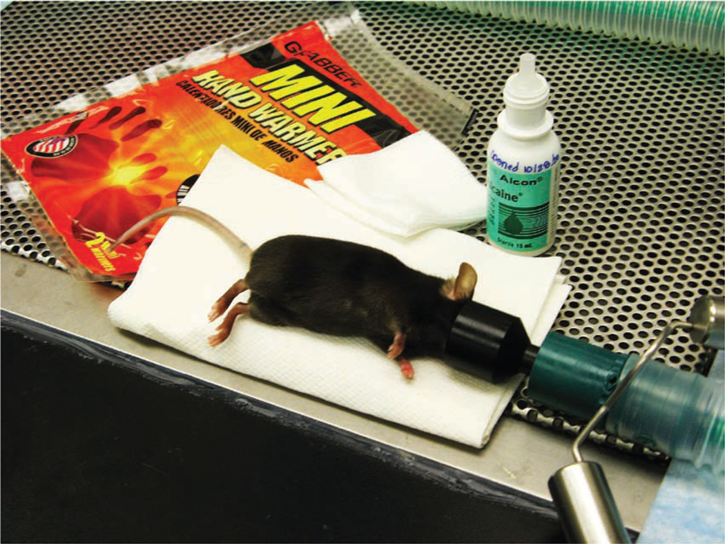
An anesthetized adult mouse placed on a protected warming device. A funnel-shaped face mask is attached to the non-rebreathing apparatus. A down-draft table is used to capture waste anesthetic gases.
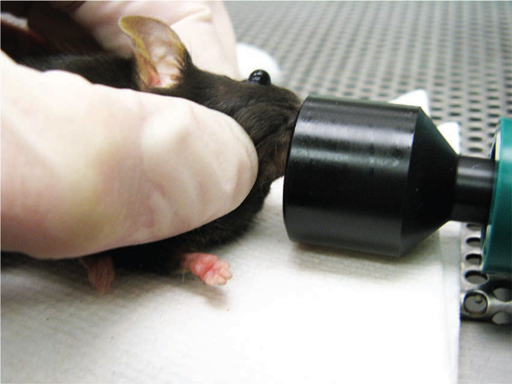
The mouse’s eye is partially protruded from the socket by applying gentle downward pressure to the skin dorsal and ventral to the eye.
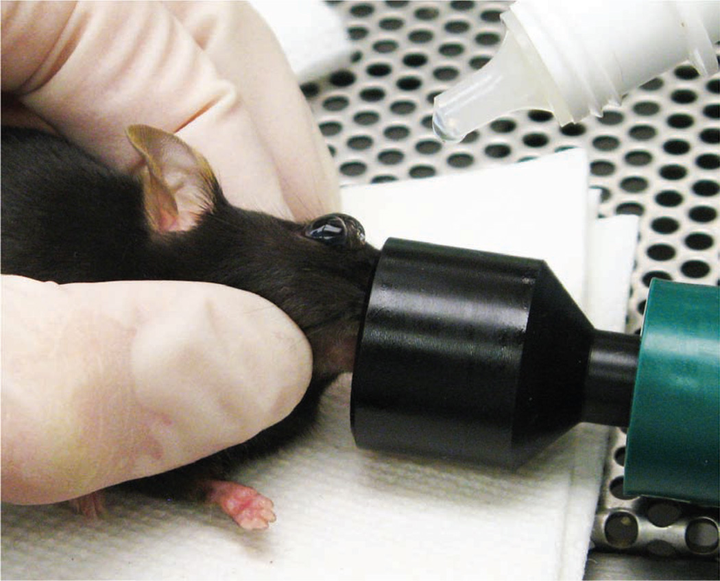
The operator places a drop of topical ophthalmic anesthetic on the eye of the mouse before carrying out the injection.
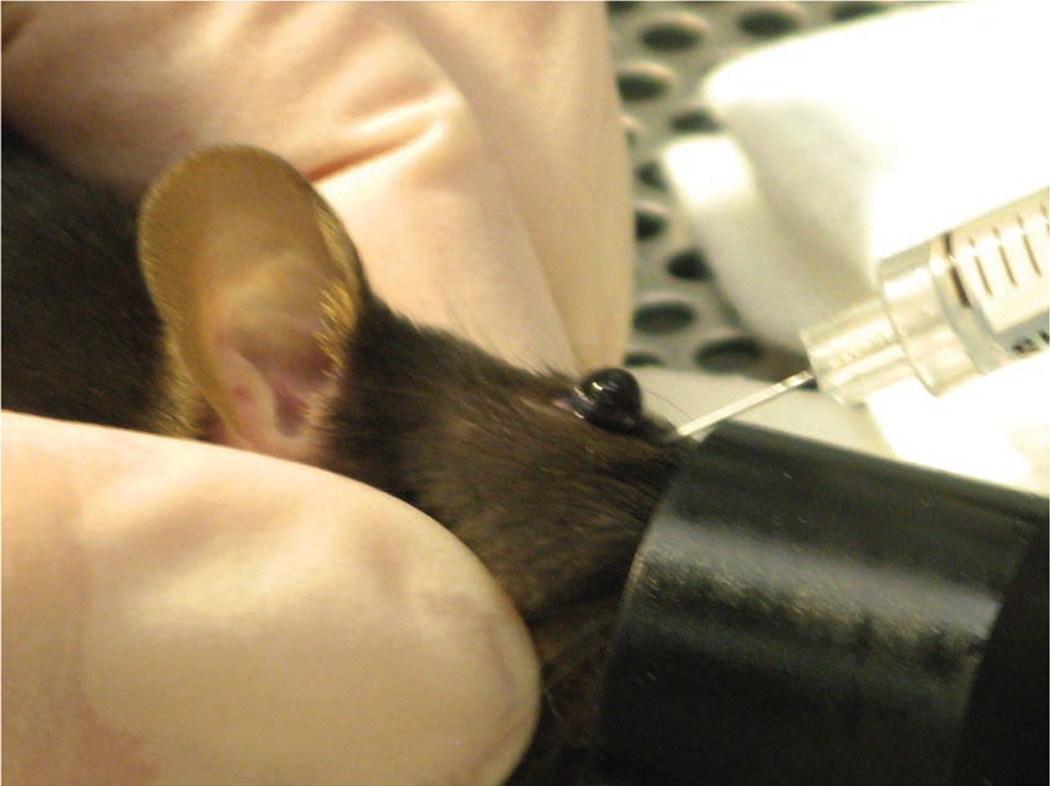
The operator inserts the needle, bevel down, at the medial canthus, into the retro-orbital sinus.
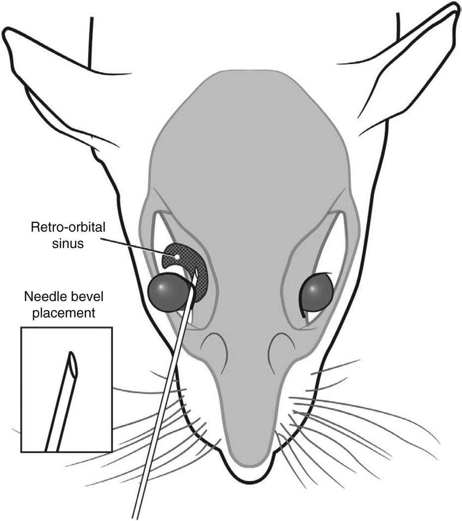
Correct placement of the needle relative to the retro-orbital sinus, the eyeball and the back of the orbit. Illustration by Darryl Leja.
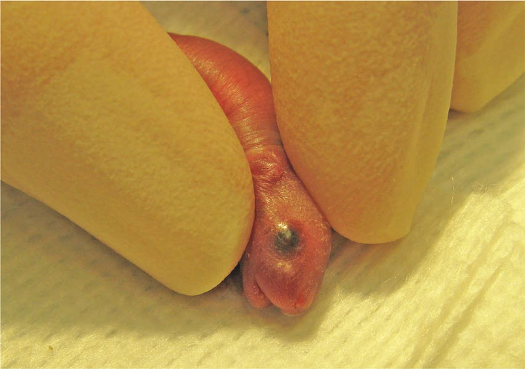
The operator gently restrains the pup between his or her thumb and forefinger.
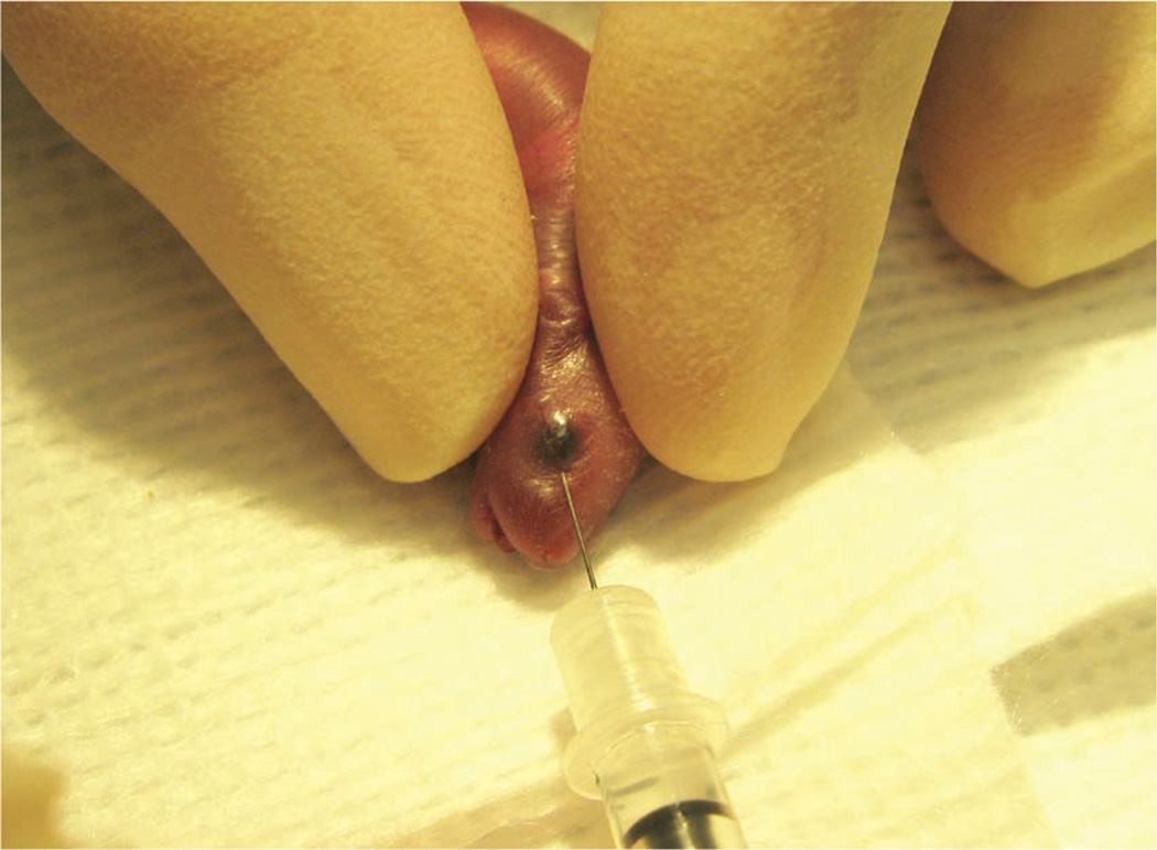
The operator inserts the needle, bevel down, at a 30° angle in the area that will become the medial canthus.
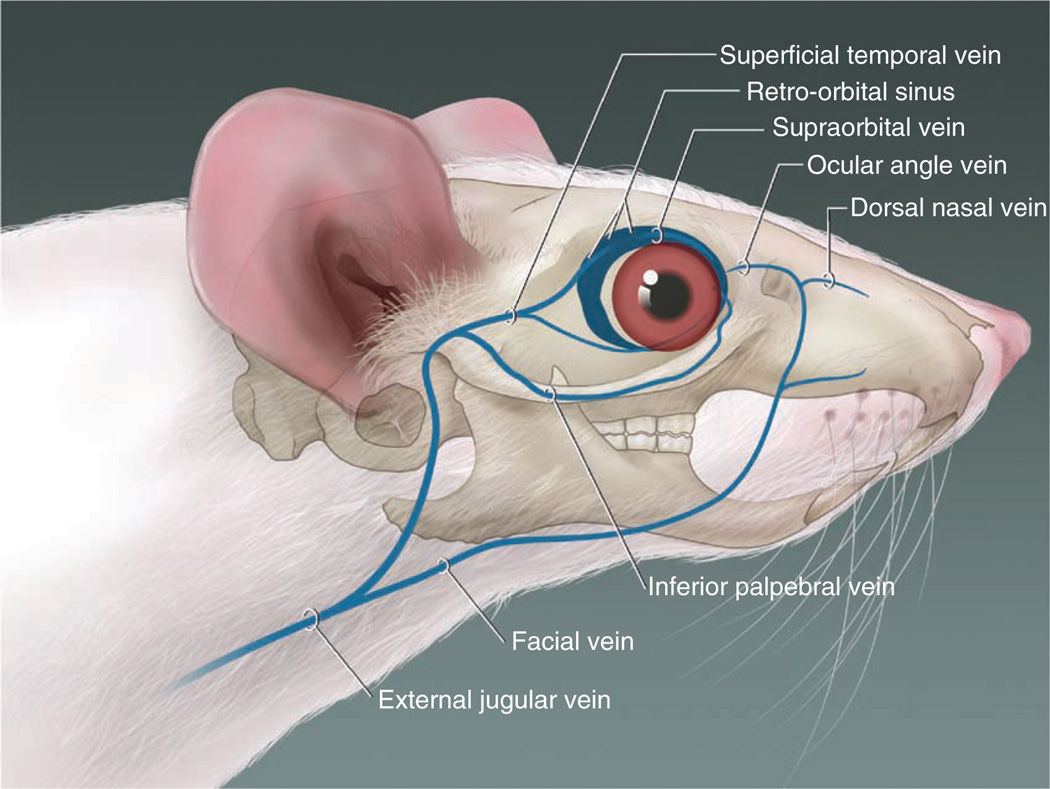
The blood vessels contributing to the retro-orbital sinus of the mouse. Illustration by Darryl Leja.
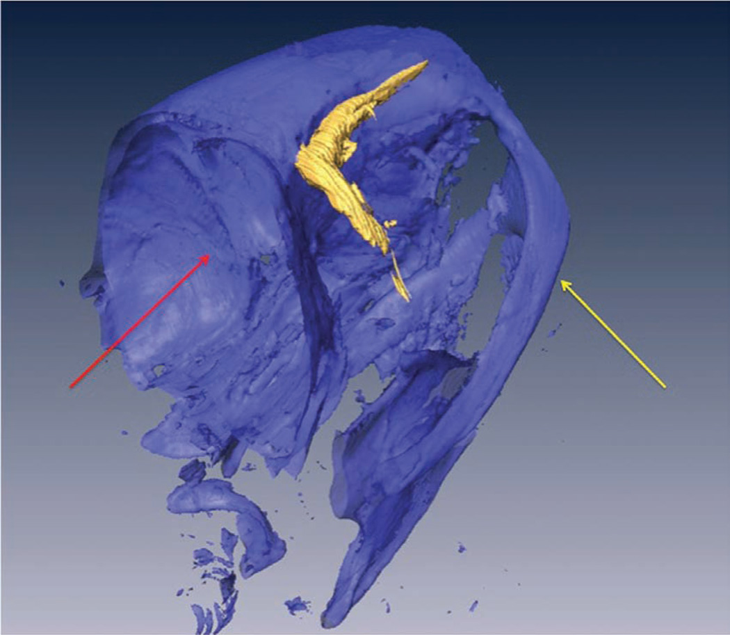
A volume-rendered three-dimensional computed tomography scan of the head of a mouse (facing right) that received a retro-orbital injection of an intravascular contrast agent. The retro-orbital sinus is in high contrast (shown in gold). The underlying bone structures are selected out with a bone-window (shown in blue) as a structural reference. The yellow arrow points to the zygomatic arch. The red arrow points to the cranial vault.
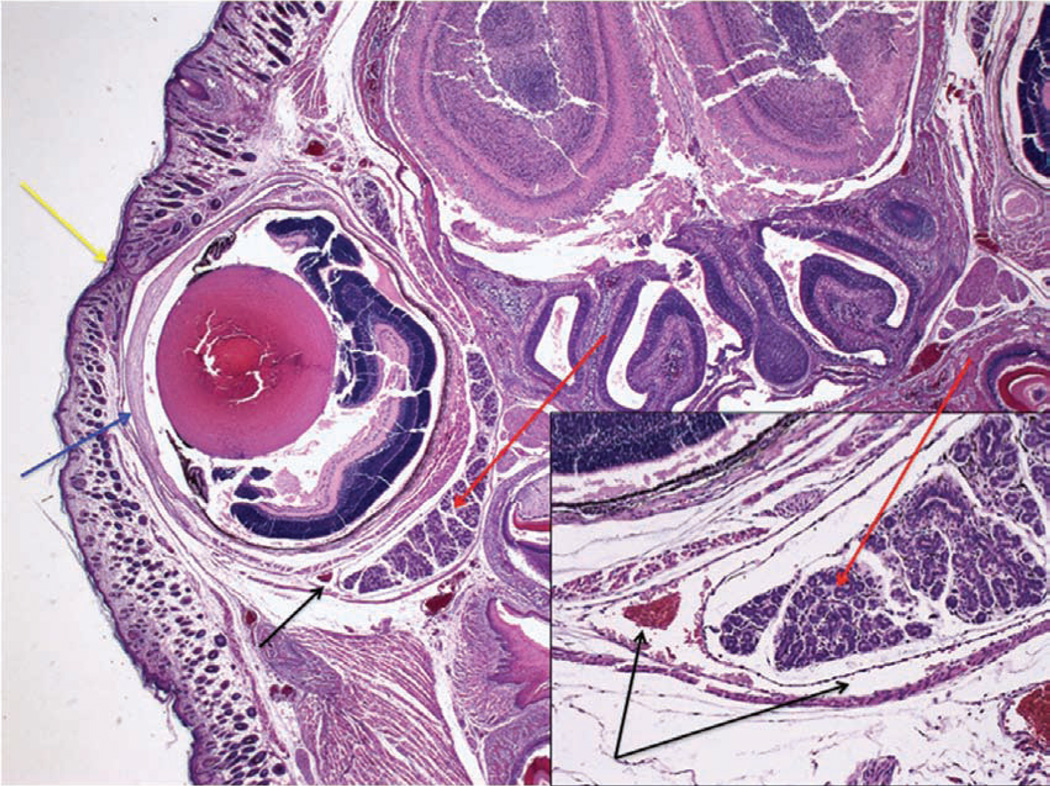
Section through the right eye and surrounding tissue of a 1-d-old mouse pup 30 min after administration of a retro-orbital injection (12.5× magnification; hematoxylin and eosin staining). The inset shows the injected retro-orbital sinus (100× magnification). The retro-orbital sinus (black arrows) and surrounding tissue appear normal. Red arrows point to the Harderian gland. Blue arrow points to the corneal surface of the eyeball. Yellow arrow points to the fused eyelids.
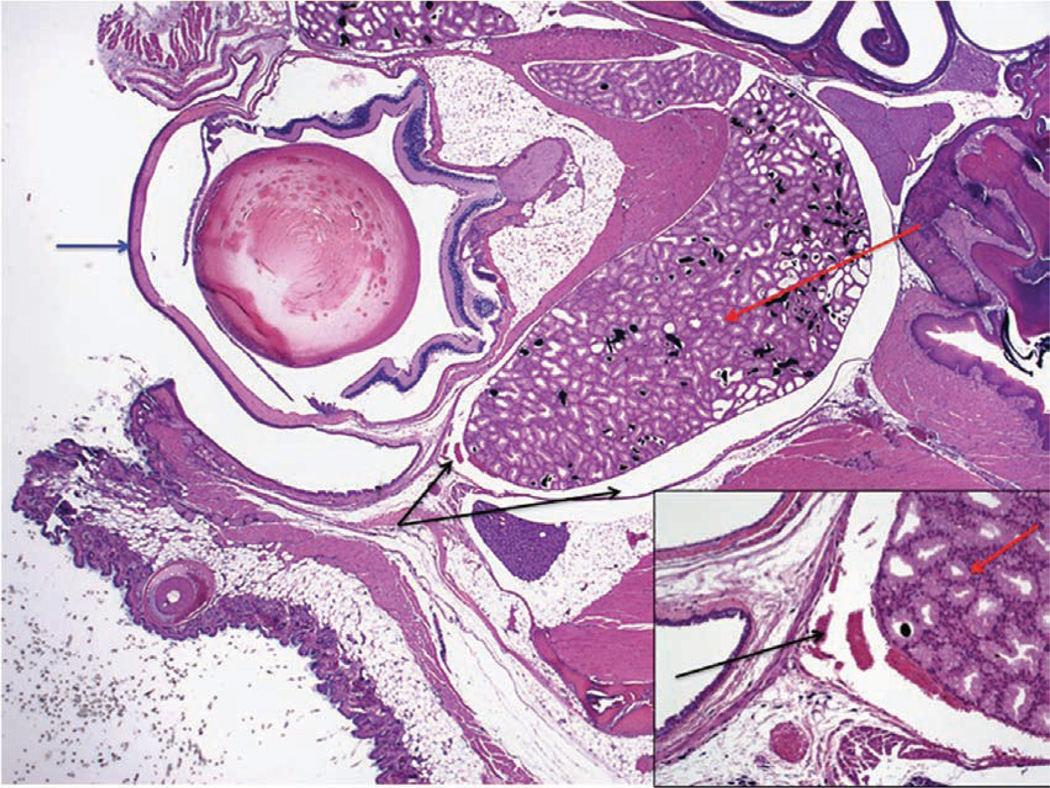
Section through the right eye and surrounding tissue of an adult mouse 30 min after administration of a retro-orbital injection (12.5× magnification; hematoxylin and eosin staining). The inset shows the injected retro-orbital sinus (100× magnification). The retro-orbital sinus (black arrows) and surrounding tissue appear normal. Red arrows point to the Harderian gland. Blue arrow points to the corneal surface of the eyeball.
References
-
- Suckow MA, Danneman P, Brayton C. In: The Laboratory Mouse. Suckow MA, editor. Roca Raton, FL: CRC; 2001.
-
- Billingham RE, Brent L. Acquired tolerance of foreign cells in newborn animals. Proc. R. Soc. Lond. B. Biol. Sci. 1956;146:78–90. - PubMed
-
- Sands MS, Barker JE. Percutaneous intravenous injection in neonatal mice. Lab. Anim. Sci. 1999;49:328–330. - PubMed
Publication types
MeSH terms
Grants and funding
LinkOut - more resources
Full Text Sources
Other Literature Sources
