Review
. 2011 May;12(5):320-32.
doi: 10.1038/nrm3107.

Centromeres: unique chromatin structures that drive chromosome segregation

Affiliations
Review

Centromeres: unique chromatin structures that drive chromosome segregation

Jolien S Verdaasdonk et al. Nat Rev Mol Cell Biol. 2011 May.

Abstract

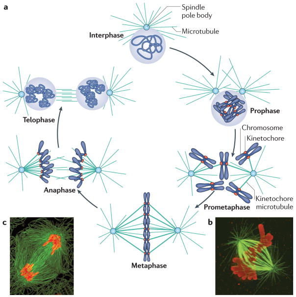
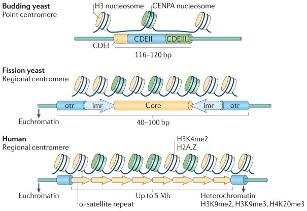
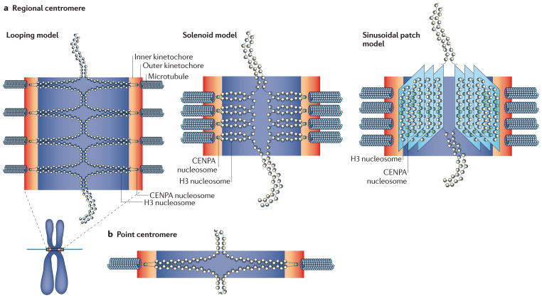
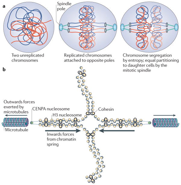
Fidelity during chromosome segregation is essential to prevent aneuploidy. The proteins and chromatin at the centromere form a unique site for kinetochore attachment and allow the cell to sense and correct errors during chromosome segregation. Centromeric chromatin is characterized by distinct chromatin organization, epigenetics, centromere-associated proteins and histone variants. These include the histone H3 variant centromeric protein A (CENPA), the composition and deposition of which have been widely investigated. Studies have examined the structural and biophysical properties of the centromere and have suggested that the centromere is not simply a 'landing pad' for kinetochore formation, but has an essential role in mitosis by assembling and directing the organization of the kinetochore.


Conflict of interest statement

Competing interests statement

The authors declare no competing financial interests.

Figures

Figure 1
Figure 1. Chromosome segregation in the cell cycle
a | The various stages of the cell cycle are depicted. During interphase, the cell undergoes growth and replication of the DNA. Upon replication of the spindle pole body and DNA, the cell undergoes a second round of growth and subsequently enters mitosis. Mitosis is divided into prophase (when the chromatin is condensed), prometaphase (when kinetochore microtubules start to interact with kinetochores), metaphase (when chromosomes become bi-oriented), anaphase (when the sister chromatids segregate to opposite spindle poles) and telophase (when chromosomes decondense). In most eukaryotes, the nuclear membrane degrades during mitosis and reforms during telophase, but this does not occur in budding yeast. b,c | Images of metaphase (b) and anaphase (c) cells. Chromosomes are shown in red and microtubules forming the mitotic spindle are shown in green. The proteinacious kinetochore forms at the centromere and mediates attachment to the spindle. Image in part b is reproduced, with permission, from REF. © (2010) The Rockefeller University Press. Image in part c is reproduced, with permission, from REF. © (2010) Macmillan Publishers Ltd. All rights reserved.
Figure 2
Figure 2. Characteristics of point and regional centromeres
The point centromeres of budding yeast form a single microtubule attachment per chromosome, whereas larger regional centromeres form multiple attachments. The budding yeast centromere DNA is composed of the conserved centromere DNA element I (CDEI), CDEII and CDEIII. Larger regional centromeres do not contain DNA sequences, but the presence of a centromeric protein A (CENPA)-containing nucleosome is conserved. H3K4me2, histone H3 dimethylated at Lys4; H3K9me, histone H3 methylated at Lys9; H3K20me3, histone H3 trimethylated at Lys20; imr, inverted repeat sequence; otr, outer repeat.
Figure 3
Figure 3. The CENPA-containing nucleosome
a | Work in different organisms on the composition and physical properties (direction of DNA wrapping and supercoiling induction) of the centromeric nucleosome has given rise to a range of different possibilities: homotypic octamer (Saccharomyces cerevisiae,, Drosophila melanogaster and human cells,,,), heterotypic octamer (human cells), reverse octamer (molecular dynamics,), homotypic tetramer or tetrasome (Schizosaccharomyces pombe) and heterotypic tetramer or hemisome (S. cerevisiae and D. melanogaster,). Furthermore, two alternative structures that contain the non-histone protein suppressor of chromosome missegregation 3 (Scm3) have been proposed: hexasomes (S. cerevisiae) and trisomes (S. cerevisiae and D. melanogaster). b | Tension on the right-handed reverse-wrapped centromeric protein A (CENPA)-containing nucleosome may cause it to split into two hemisomes (histone H2A, H2B, CENPA and H4), and could serve as a site for the spindle assembly checkpoint to monitor attachment. HJURP, Holliday junction recognition protein. Images in part a are modified, with permission, from REF. © (2011) Elsevier.
Figure 4
Figure 4. Chromatin geometry at the centromere
a | Three models for the organization of the regional centromere have been proposed: the looping model,,, the solenoid model,, and the sinusoidal patch model. The looping model proposes that the pericentric chromatin is looped out from bulk chromatin towards the spindle pole. The solenoid model proposes that the pericentric chromatin forms a coil with centromeric protein A (CENPA)-containing nucleosomes facing the spindle pole. The sinusoidal patch model attempts to explain the observed location of various constitutively centromere-associated network (CCAN) proteins and the unfolding of the vertebrate kinetochore. b | The budding yeast pericentromere adopts a cruciform structure, which serves to place the centromere (and therefore the kinetochore) on the poleward-facing side of the chromosomes. We equate the multiple loops of the looping model in part a to the whole mitotic spindle of budding yeast. H3, histone H3.
Figure 5
Figure 5. Applying the principles of polymer physics to chromosome segregation
a | Entropic forces drive the segregation of bulk chromatin. This is because it is energetically favourable for the polymers to segregate, as this allows them to adopt higher entropic states. The mitotic spindle apparatus provides directionality for this segregation and ensures that sister chromatids are equally segregated to daughter cells. b | It is important that the forces present at the mitotic spindle remain balanced to prevent breakage of the chromatin while maintaining tension along the chromatin (to sense bi-orientation). The microtubules exert an outwards force (towards the spindle pole), whereas the chromatin maintains an inwards force and is flexible enough to accommodate microtubule-based tension. CENPA, centromeric protein A; H3, histone H3.

References

    1. Moore LL, Roth MB. HCP-4, a CENP-C-like protein in Caenorhabditis elegans, is required for resolution of sister centromeres. J Cell Biol. 2001;153:1199–1208. - PMC - PubMed
    1. Sullivan BA, Karpen GH. Centromeric chromatin exhibits a histone modification pattern that is distinct from both euchromatin and heterochromatin. Nature Struct Mol Biol. 2004;11:1076–1083. - PMC - PubMed
    1. Cheeseman IM, Desai A. Molecular architecture of the kinetochore–microtubule interface. Nature Rev Mol Cell Biol. 2008;9:33–46. - PubMed
    1. Bouck DC, Joglekar AP, Bloom KS. Design features of a mitotic spindle: balancing tension and compression at a single microtubule kinetochore interface in budding yeast. Annu Rev Genet. 2008;42:335–359. - PMC - PubMed
    1. Santaguida S, Musacchio A. The life and miracles of kinetochores. EMBO J. 2009;28:2511–2531. A comprehensive review of the structure and functions of the kinetochore. - PMC - PubMed

Publication types