Review
. 2011 Aug;68(15):2513-23.
doi: 10.1007/s00018-011-0687-5. Epub 2011 Apr 21.

Regulation of self-renewal and differentiation by the intestinal stem cell niche

Affiliations
Review

Regulation of self-renewal and differentiation by the intestinal stem cell niche

Trevor M Yeung et al. Cell Mol Life Sci. 2011 Aug.

Abstract

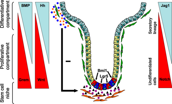
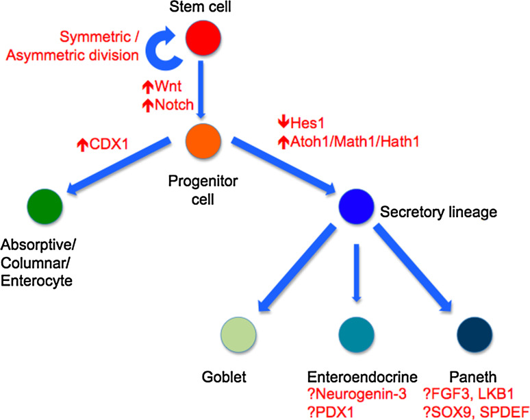
The gastrointestinal epithelium is a highly organised tissue that is constantly being renewed. In order to maintain homeostasis, the balance between intestinal stem cell (ISC) self-renewal and differentiation must be carefully regulated. In this review, we describe how the intestinal stem cell niche provides a unique environment to regulate self-renewal and differentiation of ISCs. It has traditionally been believed that the mesenchymal myofibroblasts play an important role in the crosstalk between ISCs and the niche. However, recent evidence in Drosophila and in vertebrates suggests that epithelial cells also contribute to the niche. We discuss the multiple signalling pathways that are utilised to regulate stemness within the niche, including members of the Wnt, BMP and Hedgehog pathways, and how aberrations in these signals lead to disruption of the normal crypt-villus axis. Finally, we also discuss how CDX1 and inhibition of the Notch pathway are important in specifying enterocyte and goblet cell differentiation respectively.


Figures

Fig. 1
Fig. 1
Intestinal stem cells (ISCs, red) may be located at the base of the crypt (Lgr5+ cells) or at the +4 position (Bmi1+ cells). Paneth cells (blue) are located between Lgr5+ cells. There may be overlap between the Lgr5 and Bmi1 populations. The daughter cells of ISCs migrate up the crypt–villus axis and pass through a progenitor stage, forming transient amplifying (TA) cells (yellow), before becoming differentiated cells (blue). Myofibroblasts (orange) are an integral component of the stem cell niche intimately related to the ISCs and secrete factors that regulate ISC function, including Wnt ligands (purple circles) [104] and BMP antagonists, such as Gremlin 1 (black triangles) [48]. The Wnt protein family and BMP antagonists are expressed in a reciprocal gradient to the BMP (blue circles) and Hh (yellow squares) protein families along the crypt–villus axis. Hh is expressed by differentiated colonocytes and limits expression of Wnt target genes to the base of the crypts. Activation of Hh increases BMP signalling and inhibits the Wnt pathway. Myofibroblasts are Hh responsive and regulate the patterning of the crypt–villus axis. Inhibition of the Notch pathway leads to the differentiation of ISCs into the secretory lineage. The Notch ligand Jagged-1 is expressed in a reciprocal manner to the Notch-1, -2 and -3 receptors. Non-niche intestinal stromal cells are coloured green. Small intestine epithelium contains villi, but these are absent in the large intestine. (Adapted from Kosinski et al. [48])
Fig. 2
Fig. 2
Lineage specification of an intestinal stem cell (ISC). Stem cells may divide either symmetrically or asymmetrically, although recent evidence suggest the former is most likely [14, 50]. Wnt signals maintain the stem-like phenotype of ISCs, whilst Notch maintains the proliferation of progenitor cells. The intestinal stem cell gives rise to progenitor cells, which can either form enterocytes (via CDX1) or the secretory lineage, mediated by inhibition of the Notch pathway, downregulation of Hes-1 and upregulation of Atoh1/Math1/Hath1 homologues. Paneth cells are found in the small intestine, but not normally in the large intestine apart from the proximal colon. The mechanisms that regulate specification of enteroendocrine and Paneth cells are not fully understood. Candidate genes include neurogenin-3 and PDX1 (enteroendocrine) and FGF3, LKB1, SOX9 and SPDEF (Paneth cells)

References

    1. Bjerknes M, Cheng H. Clonal analysis of mouse intestinal epithelial progenitors. Gastroenterology. 1999;116(1):7–14. doi: 10.1016/S0016-5085(99)70222-2. - DOI - PubMed
    1. Hermiston ML, Gordon JI. Organization of the crypt–villus axis and evolution of its stem cell hierarchy during intestinal development. Am J Physiol. 1995;268(5 Pt 1):G813–G822. - PubMed
    1. Wilson CL, et al. Regulation of intestinal alpha-defensin activation by the metalloproteinase matrilysin in innate host defense. Science. 1999;286(5437):113–117. doi: 10.1126/science.286.5437.113. - DOI - PubMed
    1. Ayabe T, et al. Secretion of microbicidal alpha-defensins by intestinal Paneth cells in response to bacteria. Nat Immunol. 2000;1(2):113–118. doi: 10.1038/77783. - DOI - PubMed
    1. Chang WW, Leblond CP. Renewal of the epithelium in the descending colon of the mouse. I. Presence of three cell populations: vacuolated-columnar, mucous and argentaffin. Am J Anat. 1971;131(1):73–99. doi: 10.1002/aja.1001310105. - DOI - PubMed

Publication types

MeSH terms

LinkOut - more resources