. 2011 Aug;95(8):559-71.
doi: 10.1002/bip.21638. Epub 2011 Apr 20.

Characterizing flexible and intrinsically unstructured biological macromolecules by SAS using the Porod-Debye law

Affiliations

Characterizing flexible and intrinsically unstructured biological macromolecules by SAS using the Porod-Debye law

Robert P Rambo et al. Biopolymers. 2011 Aug.

Abstract

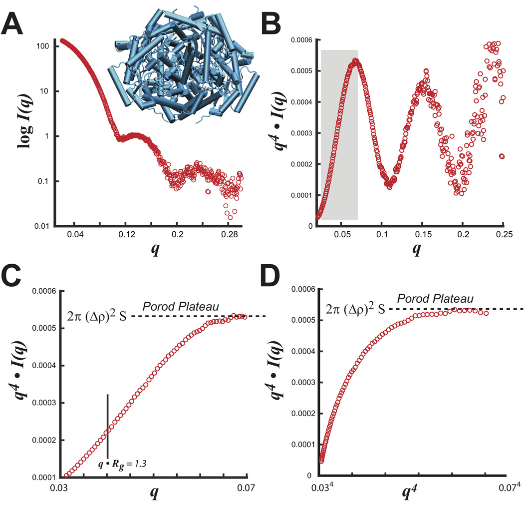
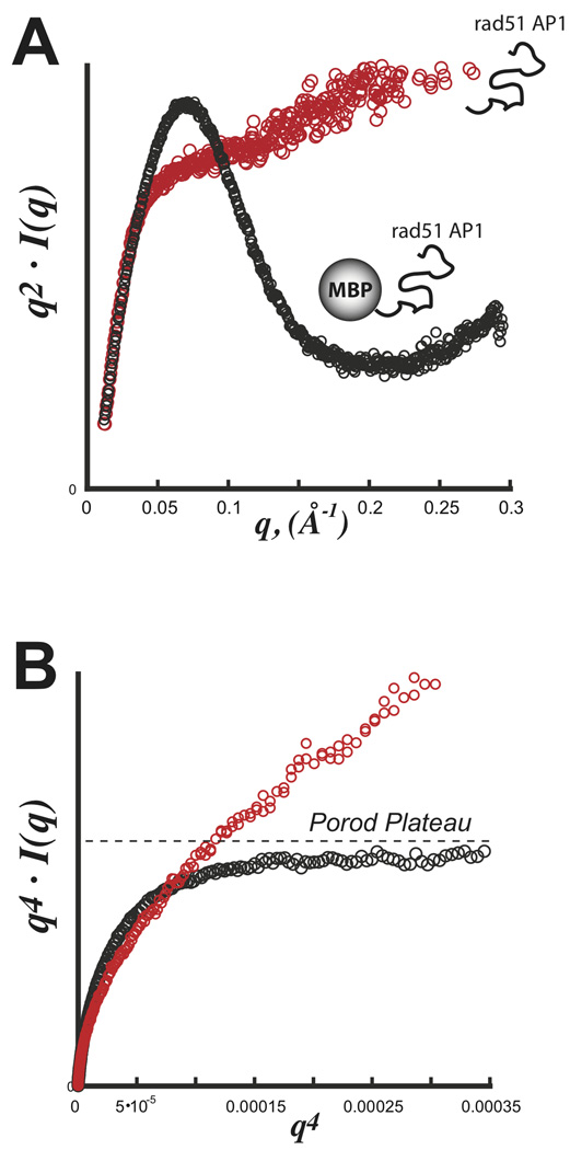
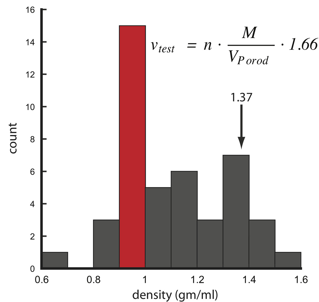
Unstructured proteins, RNA or DNA components provide functionally important flexibility that is key to many macromolecular assemblies throughout cell biology. As objective, quantitative experimental measures of flexibility and disorder in solution are limited, small angle scattering (SAS), and in particular small angle X-ray scattering (SAXS), provides a critical technology to assess macromolecular flexibility as well as shape and assembly. Here, we consider the Porod-Debye law as a powerful tool for detecting biopolymer flexibility in SAS experiments. We show that the Porod-Debye region fundamentally describes the nature of the scattering intensity decay by capturing the information needed for distinguishing between folded and flexible particles. Particularly for comparative SAS experiments, application of the law, as described here, can distinguish between discrete conformational changes and localized flexibility relevant to molecular recognition and interaction networks. This approach aids insightful analyses of fully and partly flexible macromolecules that is more robust and conclusive than traditional Kratky analyses. Furthermore, we demonstrate for prototypic SAXS data that the ability to calculate particle density by the Porod-Debye criteria, as shown here, provides an objective quality assurance parameter that may prove of general use for SAXS modeling and validation.


Figures

FIGURE 1
FIGURE 1
At low resolution (standing far from picture), differing contrasts contribute to the identification of objects within the picture. The hat, dress and grassy surroundings are clear features of the paintings with differing contrasts. Under close inspection (high resolution), only high contrast differences, such as the blue dress, allow for individual features to be distinguishable whereas differences with low contrast (boxed region) become difficult to discern. The difficulty of identifying the hat against the grassy background is analogous to the electron density contrast becoming continuous between the particle and solvent for flexible systems. This image was adapted from Matin a Eragny by Camille Pissarro and kindly prepared by Mike Pique at the Scripps Research Institute.
FIGURE 2
FIGURE 2
Canonical SAXS scattering equations. A) Guinier approximation describing the linear relationship between the observed scattering intensities, I(q), and scattering angle, q. The approximation determines the radius-of-gyration, Rg, and scattering at q=0, I(0). I(0) is directly related to the particle’s volume, V, and electron density contrast, Δρ. B) Kratky plot of scattering data illustrating changes in the behavior of the curve for folded (sphere), partially folded (sphere-random coil) and completely unfolded particles (random coil). For a folded particle, the integrated area under the curve determines the Porod invariant, Q, and is scaled by concentration, c. C) SAXS derived structural parameters. The ratio of I(0) to Q determines the volume of the scattering particle sometimes known as the Porod volume, VP. Application of the Porod-Debye law determines the surface area, S, of the scattering particle that is scaled by concentration and Δρ that can be normalized using Q.
FIGURE 3
FIGURE 3
Dimensionless Kratky analysis. A) SAXS data of a 70 kDa protein collected at two concentrations (open gray circles represent a 70% dilution of the sample in red). B) Kratky plot of the data in A where the data in gray was scaled by 1.4× dilution factor. C) Transformation equations for the dimensionless Kratky plot using Guinier parameters: Rg and I(0). D) Dimensionless Kratky transformation using the experimental Guinier parameters for each respective curve in A.
FIGURE 4
FIGURE 4
S. rubiginosus glucose isomerase. A) Experimental SAXS intensity plot with the tetrameric structure (blue). B) Porod plot of SAXS data transformed as q4 I(q) vs. q. Boxed region in gray describes the Porod-Debye region used in C and D. Examination of the boxed region in C) shows the Porod-Debye plateau that is proportional to 2π·(Δρ)2·S. D) Porod-Debye plot of SAXS data transformed as q4 I(q) vs. q4. Both plots in C and D aid in defining the Porod-Debye region for the determination of V using PRIMUS.
FIGURE 5
FIGURE 5
SAXS with an exemplary intrinsically disordered domain Rad51 AP1. Data was collected for both rad51 AP1 (red) and a fusion construct with E. coli maltose binding protein (MBP) (black). A) Kratky plot demonstrating the parabolic shape for a partially folded particle (black) and hyperbolic shape for a full unfolded particle (red). B) Porod-Debye plot demonstrating a clear plateau for the partially folded MBP-Rad51 AP1 hybrid particle. In the absence of MBP, the fully unfolded domain is devoid of any discernible plateau. Data was kindly provided by Gareth Williams at the Lawrence Berkeley National Laboratory.
FIGURE 6
FIGURE 6
Metabolite induced structural changes monitored by SAXS. A) SAXS data for the 42 kDa abscisic acid binding domain PYR1 bound (black) and apo (red). B) Porod-Debye plot demonstrating discrete plateaus in bound and apo states suggesting each state can be characterized by discretely folded particles with sharp scattering contrasts. X-ray crystal structure (PDB: 3K3K) characterizes the apo state where it is expected the loop regions reorganize collapsing the structure in the bound state. C) SAXS data for the SAM-1 riboswitch bound (black) and apo (red). Crystal structure of the bound form (blue) changes in the apo state. D) Porod-Debye plot illustrating loss of the Porod plateau in the unbound state suggesting enhanced flexibility of the riboswitch. Transforming apo SAXS data by q3 I(q) vs. q3 (inset) verifies the intensity decay is no longer q−4 but q−3. Data adapted from Nishimura, N.; Hitomi, K.; Arvai, A. S.; Rambo, R. P.; Hitomi, C.; Cutler, S. R.; Schroeder, J. I.; Getzoff, E. D. Science 2009, 326, 1373–1379.
FIGURE 7
FIGURE 7
Detecting conformational flexibility. A) SAXS data for the Mre11-Rad50 complex in the presence (black) and absence (red) of ATP. SAXS scattering profiles transformed as a Kratky plot does not confidently demonstrate flexibility (inset). B) Porod-Debye plot illustrating changes in the Porod-Debye region. Loss of the plateau suggests Mre11-Rad50 complex becomes more flexible in the absence of ATP. Transforming apo SAXS data by q3 I(q) vs. q3 (inset) verifies the intensity decay is q−3. Data was adapted from Williams, G.J., Williams, R.S., Williams, J.S., Moncalian, G., Arvai, A., Limbo, O., Guenther, G., SilDas, S., Hammel, M., Russell, P., and Tainer, J. A., (2011) ABC ATPase signature helices in Rad50 link its nucleotide state to the Mre11 interface for DNA double-strand break repair, Nature SMB, accepted.
FIGURE 8
FIGURE 8
Histogram analysis of protein densities calculated from SAXS data using 31 different proteins and conditions. For each protein, V was calculated using PRIMUS limiting the data to the Porod-Debye region. Distribution shows the most likely density is 0.9 to 1.0 gm·cm−3. The equation can be used to calculate a test density where n is the number of subunits in multimeric complexes. For hetero-complexes, n is 1. Units of M, VPorod and 1.66 are in Da, Å3 and Å3·gm·cm−3·Da−1, respectively. SAXS data was taken from http://www.bioisis.net.

References

    1. Rambo RP, Tainer JA. Curr Opin Struct Biol. 2010;20:128–137. - PMC - PubMed
    1. Dyson HJ, Wright PE. Nat Rev Mol Cell Biol. 2005;6:197–208. - PubMed
    1. Dunker AK, Silman I, Uversky VN, Sussman JL. Curr Opin Struct Biol. 2008;18:756–764. - PubMed
    1. Wells M, Tidow H, Rutherford TJ, Markwick P, Jensen MR, Mylonas E, Svergun DI, Blackledge M, Fersht AR. Proc Natl Acad Sci U S A. 2008;105:5762–5767. - PMC - PubMed
    1. Receveur V, Czjzek M, Schulein M, Panine P, Henrissat B. J Biol Chem. 2002;277:40887–40892. - PubMed

Publication types