. 2011 Aug;26(8):1891-903.
doi: 10.1002/jbmr.410.

Mechanisms inducing low bone density in Duchenne muscular dystrophy in mice and humans

Affiliations
Free PMC article

Mechanisms inducing low bone density in Duchenne muscular dystrophy in mice and humans

Anna Rufo et al. J Bone Miner Res. 2011 Aug.
Free PMC article

Abstract

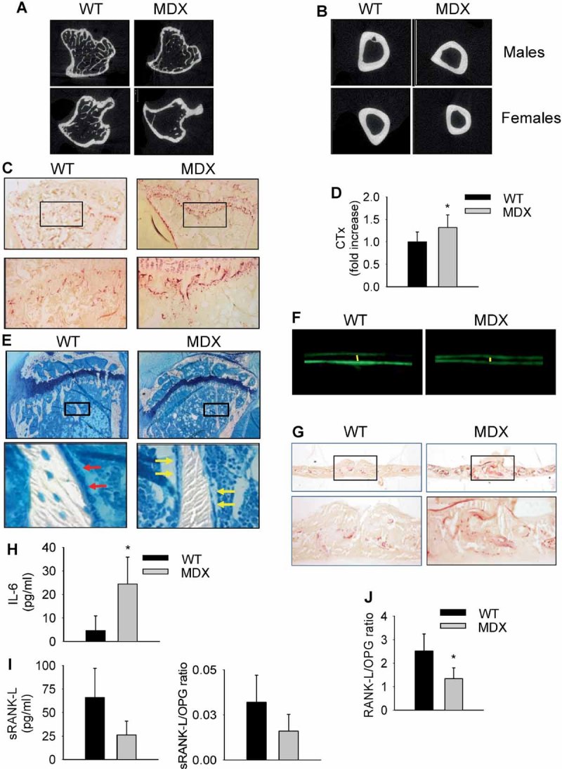
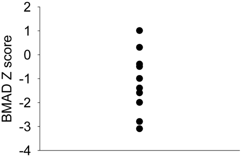
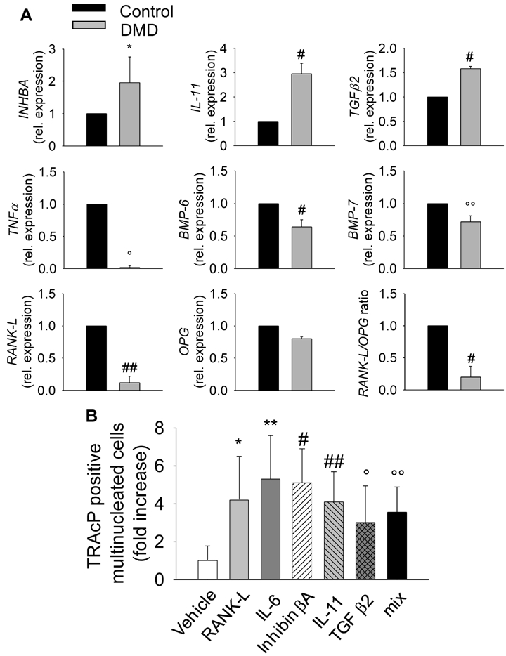
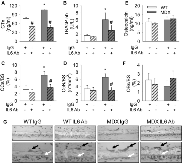
Patients affected by Duchenne muscular dystrophy (DMD) and dystrophic MDX mice were investigated in this study for their bone phenotype and systemic regulators of bone turnover. Micro-computed tomographic (µCT) and histomorphometric analyses showed reduced bone mass and higher osteoclast and bone resorption parameters in MDX mice compared with wild-type mice, whereas osteoblast parameters and mineral apposition rate were lower. In a panel of circulating pro-osteoclastogenic cytokines evaluated in the MDX sera, interleukin 6 (IL-6) was increased compared with wild-type mice. Likewise, DMD patients showed low bone mineral density (BMD) Z-scores and high bone-resorption marker and serum IL-6. Human primary osteoblasts from healthy donors incubated with 10% sera from DMD patients showed decreased nodule mineralization. Many osteogenic genes were downregulated in these cultures, including osterix and osteocalcin, by a mechanism blunted by an IL-6-neutralizing antibody. In contrast, the mRNAs of osteoclastogenic cytokines IL6, IL11, inhibin-βA, and TGFβ2 were increased, although only IL-6 was found to be high in the circulation. Consistently, enhancement of osteoclastogenesis was noted in cultures of circulating mononuclear precursors from DMD patients or from healthy donors cultured in the presence of DMD sera or IL-6. Circulating IL-6 also played a dominant role in osteoclast formation because ex vivo wild-type calvarial bones cultured with 10% sera of MDX mice showed increase osteoclast and bone-resorption parameters that were dampen by treatment with an IL-6 antibody. These results point to IL-6 as an important mediator of bone loss in DMD and suggest that targeted anti-IL-6 therapy may have a positive impact on the bone phenotype in these patients.


Figures

Fig. 1
Fig. 1
Bone phenotype in MDX mice. µCT analysis performed (A) in the tibial proximal spongiosa and (B) in cortical bone of the tibial midshafts of wild-type and MDX male and female animals. (C) Histochemical detection of the osteoclast-specific marker TRACP (purple) in proximal tibias of wild-type and MDX mice. Original magnification ×2.5 (upper panels) and ×10 (lower panels). (D) Detection of C-terminal telopeptide of type 1 collagen (CTX) in MDX sera by ELISA, as specified in “Materials and Methods.” Average value in wild-type mice was 24.5 ± 7.8 ng/mL. *p = .04 versus wild type. (E) Histologic sections of secondary proximal spongiosa of wild-type and MDX mouse proximal tibias stained with methylene blue/azure II. Original magnification ×2.5 (upper panels) and ×40 (lower panels). Red arrows = active cuboidal osteoblasts; yellow arrows = inactive flat osteoblasts. (F) Calcein (green fluorescence) labeling of secondary proximal spongiosa from wild-type and MDX mice showing the trabecular mineral apposition (distance between the two fluorescent labels, evidenced by the yellow lines). Original magnification ×40. (G) Coronal sections of calvaria from wild-type and MDX mice stained for histochemical detection of TRACP (purple). Original magnification ×2.5 (upper panels) and ×10 (lower panels). (H) Detection of IL-6 in wild-type and MDX sera by ELISA. (I) Detection of sRANKL in wild-type and MDX sera by ELISA. OPG was unchanged in the two genotypes, whereas RANK-L/OPG ratio was reduced in MDX sera and also shown in panel I. *p = .04 versus wild type. (J) RNA was extracted from femurs of 6-month-old MDX and wild-type mice and reverse transcribed; then cDNA was subjected to comparative real-time PCR using primer pairs and conditions specific for RANKL and OPG. RANK-L/OPG ratio is shown. *p = .003 versus wild type. Values are normalized versus the house keepinggene GAPDH. All values are the mean ± SD of at least four sera samples or three animals per group.
Fig. 2
Fig. 2
Distribution of the 16 DMD patients along the bone mineral apparent density (BMAD) Z-score gradient.
Fig. 3
Fig. 3
Effects of sera from DMD patients on in vitro human bone cells. (A) Osteoblasts were cultured for 3 weeks in the presence of 10% sera pooled from healthy donors (Control) or DMD patients and with ascorbic acid and β-glycerophosphate, as described in “Materials and Methods,” to favor mineralization. Cultures then were fixed and stained to reveal mineralized nodules by the von Kossa reaction (dark staining, panels). Intensity of mineralization was measured by densitometry (graphs). *p = .0005 versus control. (B) Osteoblasts were grown as above and then evaluated for alkaline phosphatase (ALP) activity by histochemical detection (panels, dark staining) or by biochemical detection (graphs). (C) Osteoblasts were grown in the presence of 10% sera pooled from healthy controls or DMD patients, with an IL-6-blocking antibody (IL-6 Ab) or an irrelevant IgG, as indicated. After 48 hours, RNA was extracted and reverse transcribed, and cDNA was subjected to comparative real-time PCR using primer pairs and conditions specific for osterix (OSX), osteocalcin (OCN) and Runt-related transcription factor 2 (RUNX2). #p = .004 and *p = .0005 versus control. (D) Human peripheral blood mononuclear cells from healthy donors were cultured in the presence of 25 ng/mL of M-CSF, 0.5 ng/mL of sRANKL, and 10% sera from healthy donors (Control) or DMD patients and evaluated for TRACP positivity, as described in “Materials and Methods.” *p = .003 versus control. (E) Human peripheral blood mononuclear cells from control subjects or 3 DMD patients were cultured in the presence of 10% FBS, 25 ng/mL of M-CSF, and 30 ng/mL of sRANKL and evaluated for TRACP positivity, as described in “Materials and Methods.” #p = .05 versus control. Average numbers of TRACP+ multinucleated osteoclasts under control conditions was 29.8 ± 19.4/well in panel D and 36.0 ± 10.2/well in panel E. All values are the mean ± SD of at least three independent experiments. Original magnification ×10.
Fig. 4
Fig. 4
Cytokine expression and osteoclastogenesis assay. (A) Osteoblasts from healthy donors were incubated with sera from controls or DMD patients, as described in “Materials and Methods.” RNA was extracted and reverse transcribed, and cDNA was subjected to comparative real-time PCR using primer pairs and conditions specific for inhibin-βA (INHBA), IL11, TGFβ2, TNFα, bone morphogenic protein 6 (BMP6), BMP7, RANKL, and OPG. RANKL/OPG ratio is also shown. #p = .02; °°p = .03; *p = .04; °p = .002; and ##p = .003 versus control. Values are normalized versus the housekeeping gene GAPDH. (B) Human peripheral blood mononuclear cells from healthy donors were cultured, as described in “Materials and Methods,” in the presence of M-CSF and 10 ng/mL of IL-6, IL-11, inhibin-βA, TGF-β2, or sRANKL as positive control. TRACP+ multinucleated cells were enumerated and expressed as fold increase of control. Average numbers of osteoclasts in control conditions (Vehicle) was 19.0 ± 6.0/well. **p = .02; *p = .04; #p = .007; ##p = .003; and °p = .0007 versus control. All values are the mean ± SD of three independent experiments.
Fig. 5
Fig. 5
Effect of blocking IL-6 activity on murine calvarial bones in culture. Four-day-old CD1 mouse calvarial bones were cultured as described in “Materials and Methods” in the presence of a blocking antibody against IL-6 (IL-6 Ab) or an irrelevant IgG. ELISA assay for detection of (A) C-terminal telopeptide of type 1 collagen (CTX) and (B) TRACP-5b in conditioned medium. Histomorphometric evaluation of (C) osteoclast surface/bone surface (OcS/BS, %) and (D) osteoclast number/bone surface (OcN/BS, n/mm2). (E) ELISA assay for osteocalcin in conditioned medium. (F) Histomorphometric evaluation of osteoblast surface/bone surface (ObS/BS, %). (G) Coronal semithin sections of the calvarial bones stained with methylene blue/azure II. Original magnification ×20 (upper panels) and ×40 (lower panels). Black arrows = osteoclasts; white arrows = osteoblasts. Values are the mean ± SD of three calvarial bones per group.*p = .04 versus wild type with irrelevant IgG; #p = .03 versus each group treated with irrelevant IgG.

References

    1. Kunkel LM, Beggs AH, Hoffman EP. Molecular genetics of Duchenne and Becker muscular dystrophy: emphasis on improved diagnosis. Clin Chem. 1989;35:B21–B24. - PubMed
    1. Emery AE. Population frequencies of inherited neuromuscular diseases: a world survey. Neuromusc Disord. 1991;1:19–29. - PubMed
    1. Suresh S, Wales P, Dakin C, Harris MA, Cooper DG. Sleep-related breathing disorder in Duchenne muscular dystrophy: disease spectrum in the paediatric population. J Paediatr Child Health. 2005;41:500–503. - PubMed
    1. Ervasti JM, Campbell KP. A role for distrohyn-glycoprotein complex as an transmembrane linker between laminin and actin. J Cell Biol. 1993;122:809–823. - PMC - PubMed
    1. Campbell KP. Three muscular dystrophies: loss of cytoskeleton-extracellular matrix linkage. Cell. 1995;80:675–679. - PubMed

Publication types

MeSH terms