. 2011 May;33(5):1184-93.
doi: 10.1002/jmri.22530.

Adaptive noise cancellation to suppress electrocardiography artifacts during real-time interventional MRI

Affiliations

Adaptive noise cancellation to suppress electrocardiography artifacts during real-time interventional MRI

Vincent Wu et al. J Magn Reson Imaging. 2011 May.

Abstract

Purpose: To develop a system for artifact suppression in electrocardiogram (ECG) recordings obtained during interventional real-time magnetic resonance imaging (MRI).

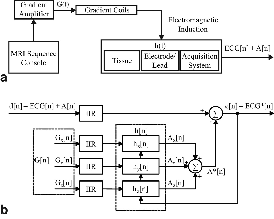
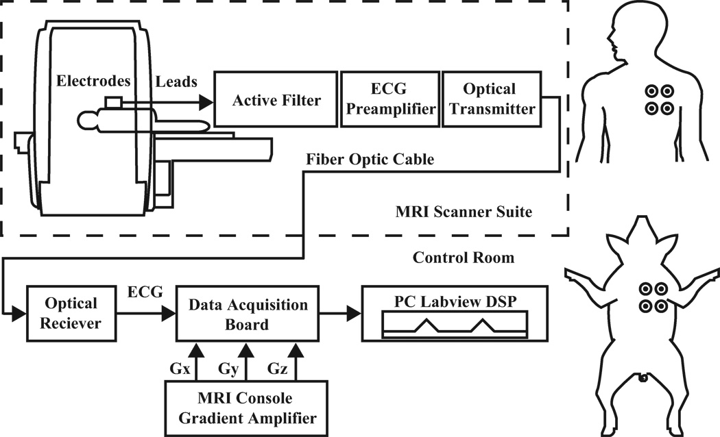
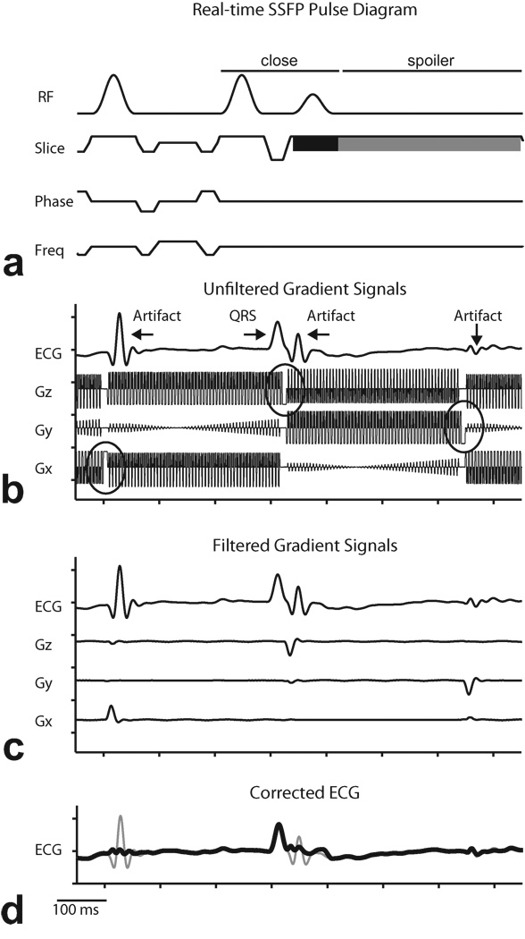
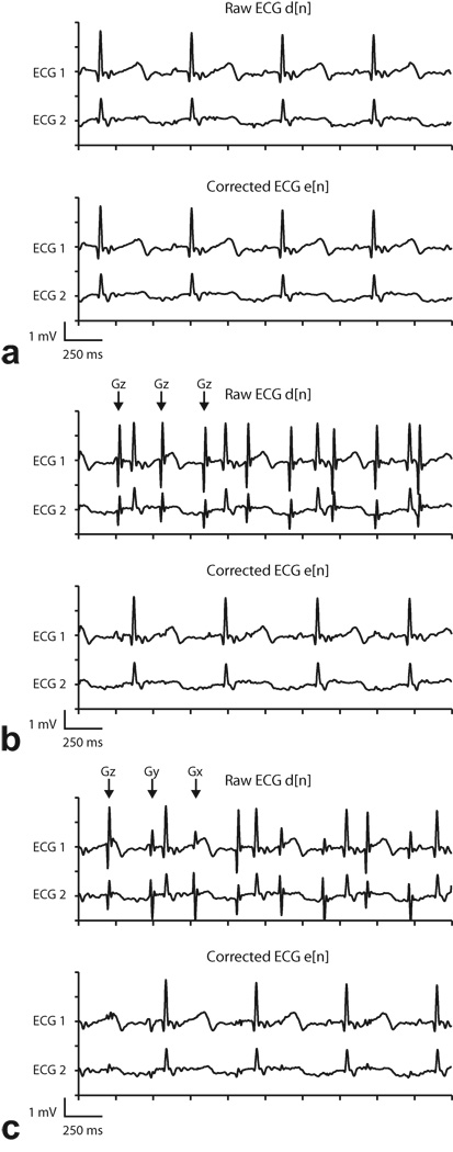
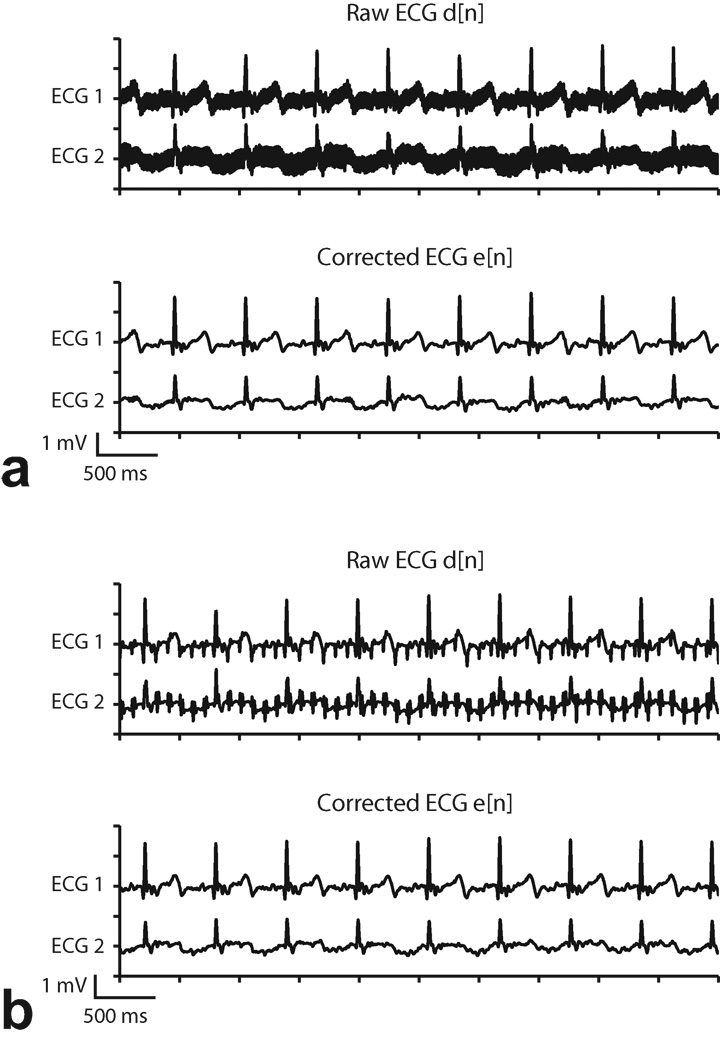
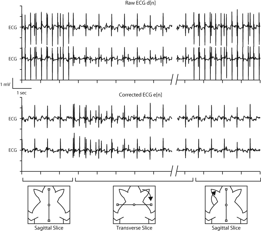
Materials and methods: We characterized ECG artifacts due to radiofrequency pulses and gradient switching during MRI in terms of frequency content. A combination of analog filters and digital least mean squares adaptive filters were used to filter the ECG during in vivo experiments and the results were compared with those obtained with simple low-pass filtering. The system performance was evaluated in terms of artifact suppression and ability to identify arrhythmias during real-time MRI.

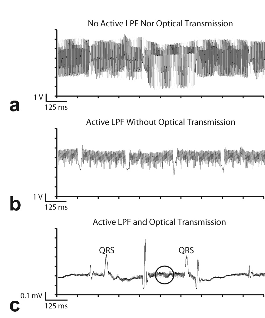
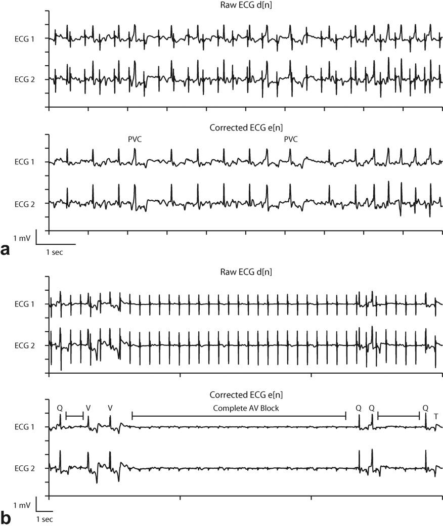
Results: Analog filters were able to suppress artifacts from high-frequency radiofrequency pulses and gradient switching. The remaining pulse artifacts caused by intermittent preparation sequences or spoiler gradients required adaptive filtering because their bandwidth overlapped with that of the ECG. Using analog and adaptive filtering, a mean improvement of 38 dB (n = 11, peak QRS signal to pulse artifact noise) was achieved. This filtering system was successful in removing pulse artifacts that obscured arrhythmias such as premature ventricular complexes and complete atrioventricular block.

Conclusion: We have developed an online ECG monitoring system employing digital adaptive filters that enables the identification of cardiac arrhythmias during real-time MRI-guided interventions.


Figures

Figure 1
Figure 1
a: Linear shift invariant modeling of Faraday induction by MRI gradient switching. b: Architecture of least mean squares adaptive filtering.
Figure 2
Figure 2
Left: Diagram of ECG instrumentation and acquisition setup. Right: Subject electrode placements.
Figure 3
Figure 3
Example of ECG artifacts during real-time SSFP. a: Results from acquisition using coaxial cable transmission lines without 4-pole analog low pass filter in the ECG sensor. b: 4-pole analog low pass filters partially suppress MRI ECG artifacts. Coaxial cables are still vulnerable to RF induction. c: ECG SNR improved with optical system and analog filter. Some residual RF artifact remains (circle).
Figure 4
Figure 4
Demonstration of digital preprocessing of reference signals in least mean squares adaptive filtering. a: Pulse sequence diagram for multi-slice real-time SSFP showing closing sequence to store magnetization (“close”), combined slice select (black) and spoiler (gray) on slice gradient. b: Example of low-pass filtered contaminated ECG recorded simultaneously with gradient signals acquired from scanner during multi-slice real-time SSFP imaging. Circles denote spoiler gradients. c: Magnified plot showing relationship between spoiler gradients and ECG pulse artifacts. d: Corrected ECG (thick line) overlaid on ECG signal before adaptive filtering (thin line).
Figure 5
Figure 5
Representative two channel ECG recordings and adaptive filtering results during real-time SSFP (Subject 1). “Raw ECG” signal includes analog low-pass filtering but not adaptive filtering, and “Corrected ECG” signal includes analog and adaptive filtering. Arrows indicate the gradient axis responsible for the spoiler gradient artifact. a: One slice (no spoilers). Gradient artifacts were suppressed sufficiently by low-pass filtering, and adaptive filter did not introduce any additional artifacts. b: One slice with fat saturation. Large Gz gradient artifacts (which occur once per slice, or every ~350 ms)are evident in low-pass filtered signal but are suppressed by adaptive filter. c: Three slices. Gradient artifacts appear on each axis, once per slice. Largest amplitude artifacts are from Gz, but artifacts from Gx and Gy are also evident in Raw ECG signal. Artifacts from all three axes are adequately suppressed by adaptive filter.
Figure 6
Figure 6
Adaptive filter results during GRE (Subject 1). a: TR= 14 ms. b: TR = 100 ms. “Raw ECG” signal includes analog low-pass filtering but not adaptive filtering, and “Corrected ECG” signal includes analog and adaptive filtering. Gradient switching artifacts are evident in the Raw ECG signal and are not eliminated by the analog low-pass filter, but are significantly suppressed by adaptive filtering.
Figure 7
Figure 7
Example of least mean squares coefficients update during interactive real-time MRI. Upon changing slice orientation from sagittal to transverse, the resulting change in pulse artifacts causes update of filter coefficients (from an initialized zero value). After approximately 4 seconds, denoised ECG reaches steady state and adapts to the new sequence settings. When the slice returns to sagittal orientation, the filter coefficients require little or no updating, and the ECG remains clean.
Figure 8
Figure 8
Adaptive filter results during arrhythmia induction. Identification of premature ventricular contractions (PVC) (a) and transient complete atrioventricular (AV) block (b) was enhanced in the corrected ECG waveforms. In (b), Q=normal QRS, V=ventricular escape beat, T=T wave, and |-|= interval of complete atrioventricular block. See text for details.

References

    1. Saikus C, Lederman R. Interventional Cardiovascular Magnetic Resonance Imaging: A New Opportunity for Image-Guided Interventions. JACC Cardiovascular Imaging. 2009;2(11):1321. - PMC - PubMed
    1. Birkholz T, Schmid M, Nimsky C, Schüttler J, Schmitz B. ECG artifacts during intraoperative high-field MRI scanning. Journal of Neurosurgical Anesthesiology. 2004;16(4):271. - PubMed
    1. Felblinger J, Lehmann C, Boesch C. Electrocardiogram acquisition during MR examinations for patient monitoring and sequence triggering. Magnetic Resonance in Medicine. 1994;32(4):523–529. - PubMed
    1. Allen P, Polizzi G, Krakow K, Fish D, Lemieux L. Identification of EEG events in the MR scanner: the problem of pulse artifact and a method for its subtraction. Neuroimage. 1998;8(3):229–239. - PubMed
    1. Nijm G, Swiryn S, Larson A, Sahakian A. Extraction of the magnetohydrodynamic blood flow potential from the surface electrocardiogram in magnetic resonance imaging. Medical and Biological Engineering and Computing. 2008;46(7):729–733. - PubMed

Publication types