. 2011 Apr 21;13(2):R46.
doi: 10.1186/bcr2868.

Trastuzumab-DM1 causes tumour growth inhibition by mitotic catastrophe in trastuzumab-resistant breast cancer cells in vivo

Affiliations

Trastuzumab-DM1 causes tumour growth inhibition by mitotic catastrophe in trastuzumab-resistant breast cancer cells in vivo

Mark Barok et al. Breast Cancer Res. .

Abstract

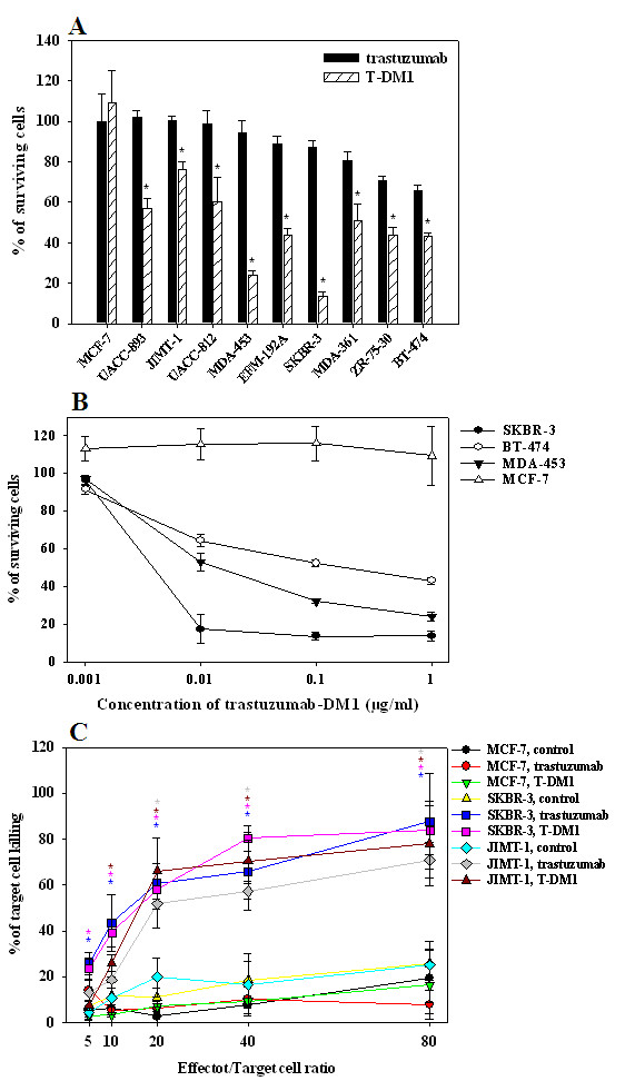
Introduction: Trastuzumab is widely used for the treatment of HER2-positive breast cancer. Despite encouraging clinical results, a significant fraction of patients are, or become, refractory to the drug. To overcome this, trastuzumab-DM1 (T-DM1), a newer, more potent drug has been introduced. We tested the efficacy and mechanisms of action of T-DM1 in nine HER2-positive breast cancer cell lines in vitro and in vivo. The nine cell lines studied included UACC-893, MDA-453 and JIMT-1, which are resistant to both trastuzumab and lapatinib.

Methods: AlamarBlue cell-proliferation assay was used to determine the growth response of breast cancer cell lines to trastuzumab and T-DM1 in vitro. Trastuzumab- and T-DM1-mediated antibody-dependent cellular cytotoxicity (ADCC) was analysed by measuring the lactate dehydrogenase released from the cancer cells as a result of ADCC activity of peripheral blood mononuclear cells. Severe Combined Immunodeficient (SCID) mice were inoculated with trastuzumab-resistant JIMT-1 cells to investigate the tumour inhibitory effect of T-DM1 in vivo. The xenograft samples were investigated using histology and immunohistochemistry.

Results: T-DM1 was strongly growth inhibitory on all investigated HER2-positive breast cancer cell lines in vitro. T-DM1 also evoked antibody-dependent cellular cytotoxicity (ADCC) similar to that of trastuzumab. Outgrowth of JIMT-1 xenograft tumours in SCID mice was significantly inhibited by T-DM1. Histologically, the cellular response to T-DM1 consisted of apoptosis and mitotic catastrophe, the latter evidenced by an increased number of cells with aberrant mitotic figures and giant multinucleated cells.

Conclusions: Our results suggest mitotic catastrophe as a previously undescribed mechanism of action of T-DM1. T-DM1 was found effective even on breast cancer cell lines with moderate HER2 expression levels and cross-resistance to trastuzumab and lapatinib (MDA-453 and JIMT-1).


Figures

Figure 1
Figure 1
In vitro effects of trastuzumab and T-DM1. A. Growth inhibitory effects of trastuzumab and T-DM1 on HER2 positive breast cancer cell lines in vitro. T-DM1 (1 μg/ml, hatched column) significantly inhibited the growth of all HER2 positive breast cancer cell lines in comparison to trastuzumab (10 μg/ml, black column) (*, P < 0.05). Neither trastuzumab nor T-DM1 had any growth inhibitory effect on MCF-7 cells with low trastuzumab binding capacity. Note that trastuzumab was used in a 10-times higher dose than T-DM1. B. T-DM1 inhibited the growth of HER2 positive breast cancer cells in a dose-dependent manner. The effect of T-DM1 was tested at a concentration of 0.001, 0.01, 0.1 and 1 μg/ml. Its dose-dependent growth inhibitory effect was seen on SKBR-3 (black circle), BT-474 (white circle) and MDA-453 (black triangle) HER2 positive breast cancer cell lines. No inhibition of HER2 negative MCF-7 (white triangle) breast cancer cells was seen. C. Trastuzumab and T-DM1 evoke similar efficient ADCC on HER2 positive breast cancer cells in vitro. Target tumour cells (SKBR-3, JIMT-1, MCF-7) were mixed with peripheral blood mononuclear cells (PBMCs) freshly isolated from peripheral blood at effector/target ratios of 5:1, 10:1, 20:1, 40:1, and 80:1. ADCC was analyzed by measuring the lactate dehydrogenase (LDH) released from the cancer cells as a result of ADCC activity of PBMCs in the presence of trastuzumab, T-DM1 or control antibody (rituximab) (20 μg/ml). The percentage of killed cells was calculated as described in Materials and methods. Killing of SKBR-3 cells was significantly higher in the presence of both trastuzumab (blue) and T-DM1 (pink) in comparison to that in the presence of control antibody (yellow) when the effector/target cell ratio was 5 or above (blue and pink stars, P < 0.05). Similarly, significantly higher killing of JIMT-1 cells was detected in the presence of both trastuzumab (gray) and T-DM1 (brown) compared to that in the presence of control antibody (pale blue) when the effector/target cell ratio was 20 or above (gray and brown stars, P < 0.05). Trastuzumab and T-DM1 evoked similar tumour cell killing on the HER2 positive breast cancer cell lines (SKBR-3 and JIMT-1). MCF-7 cells with weak trastuzumab binding capacity showed low levels of both trastuzumab- and T-DM1-evoked ADCC-mediated killing (red and green, respectively).
Figure 2
Figure 2
Correlations of drug responses and trastuzumab binding capacities. A. Correlation of response to trastuzumab and trastuzumab binding capacity. No significant correlation was found when trastuzumab binding capacities (determined by flow cytometry [40]) were plotted against trastuzumab sensitivities of the breast cancer cell lines (R = 0.43, P = 0.19). Trastuzumab sensitivities are expressed in proportion to the BT-474 cell line. B. Correlation of response to T-DM1 and trastuzumab binding capacity. No significant correlation was found when trastuzumab binding capacities were plotted against T-DM1 sensitivities of the breast cancer cell lines (R = 0.58, P = 0.06). T-DM1 sensitivities are expressed in proportion to the BT-474 cell line. C. Correlation of trastuzumab and T-DM1 responses. Effects of both drugs on breast cancer cell lines were plotted. No significant correlation was found between the responsiveness to trastuzumab and T-DM1 (R = 0.6, P = 0.07). Drug sensitivities are expressed in proportion to the BT-474 cell line.
Figure 3
Figure 3
Effect of T-DM1 on the growth of JIMT-1 xenografts. SCID mice were injected s.c. with 5 × 10⁶ JIMT-1 cells. Tumours were formed in all mice in 11 days (n = 18, mean tumour volume 64 ± 9 mm3). Thereafter, weekly treatments (arrows) with control antibody (rituximab, 5 mg/kg, i.p., white circle, n = 6), trastuzumab (5 mg/kg, i.p., black triangle, n = 6) or T-DM1 (15 mg/kg, i.v., white triangle, n = 6) were started on Day 12 and continued until the end of the experiment. A partial but significant tumour growth inhibition was seen by T-DM1 from days 32 to 44 (*, P < 0.05), while trastuzumab had no inhibitory effect on tumour growth.
Figure 4
Figure 4
Effect of T-DM1 on the formation of JIMT-1 xenografts. SCID mice injected s.c. with 5 × 10⁶ JIMT-1 cells were treated on a weekly basis with control antibody (rituximab, 5 mg/kg, i.p., black circle, n = 7), trastuzumab (5 mg/kg, i.p., red triangle, n = 6), T-DM1 (5 mg/kg, i.v., green triangle, n = 6) or on a daily basis with lapatinib (100 mg/kg, p.o., brown circle, n = 7) starting from the day of tumour inoculation (Day 0). Drug administration schedule is shown by arrows. Lapatinib (brown circle) had no effect on tumour growth. Trastuzumab (red triangle) reduced the formation of tumours between days 6 and 34 compare to both control antibody and lapatinib (red stars, P < 0.05). In three out of the six trastuzumab treated mice, trastuzumab administration was suspended and switched to T-DM1 (5 mg/kg, i.v., yellow square) from the Day 41 (marked by white arrow) whereas it was continued for another three weeks in the other three (5 mg/kg, i.p., blue square). In this setting, T-DM1 was unable to inhibit tumour growth. The tumours started to grow in four T-DM1 treated mice (green triangle) from Day 55 onward, but remained non-palpable in two (2/6). T-DM1 administration of these two mice was suspended on Day 76 (marked by Ο) and the mice were followed up for more four weeks (pale blue square, n = 2). In these two mice, a very small palpable tumour (approximately 4 mm3) was detected in one mouse on Day 107 and residual cancer cells were found histologically. No tumour was detected in the other mouse and the histological examination of the tumour inoculation site suggested a complete cure. T-DM1 treatment of the other four mice was continued until the end of the experiment (5 mg/kg, i.v., pink square, n = 4). Overall, T-DM1 significantly inhibited the tumour formation from Day 13 to Day 34 in comparison to both control antibody and lapatinib (green stars, P < 0.05), and it had a significantly growth inhibition between days 13 and 62 compared to trastuzumab (green a, P < 0.05). Note that both trastuzumab and T-DM1 were used in a dose of 5 mg/kg.
Figure 5
Figure 5
T-DM1 induced mitotic catastrophe and apoptosis in JIMT-1 xenografts. For histology, JIMT-1 tumours were obtained from SCID mice treated with control antibody (rituximab, black column), lapatinib (gray column), trastuzumab (white column), T-DM1 (hatched column) or T-DM1 after trastuzumab (cross-hatched column). Histological sections were stained with hematoxylin and eosin. When enumerating the number of mitoses with normal morphology, no differences were found between the samples (A). In contrast, a significantly higher number of cells with aberrant mitotic morphology was detected in T-DM1 treated tumours (B) (*, P < 0.05). The number of giant multinucleated cells was also increased in T-DM1 treated samples (C) (*, P < 0.05). Notably, higher number of cells with aberrant mitotic morphology and higher number of giant multinucleated cells were detected in the samples whose trastuzumab treatment was changed to T-DM1 (B-C) (*, P < 0.05). CytoDeath staining was used to detect the apoptotic cells in the histological sections. Significantly increased number of apoptotic cells was found in the T-DM1 treated samples and also in the in the samples whose trastuzumab treatment were changed to T-DM1 (D) (*, P < 0.05). Note that CytoDeath positive cells are plotted on different scales.
Figure 6
Figure 6
Histological characterization of T-DM1 induced changes in JIMT-1 xenografts. JIMT-1 xenografts were obtained from mice treated with trastuzumab (A, C, E) or T-DM1 (B, D, F). Histological sections were stained with hematoxylin and eosin (H&E, A, B). HER2 protein was visualized by HercepTest (C-D). Apoptotic cells were visualized by M30 CytoDeath antibody (E-F). High number of cells with aberrant mitotic shape (yellow arrow) and giant multinucleated cells (green arrow), which are the hallmarks of mitotic catastrophe, were seen in T-DM1 treated samples (B, D, F). Furthermore, higher number of apoptotic cells was found in the T-DM1 treated samples than in the trastuzumab treated ones (white arrow, E and F, respectively). Similar number of cells with normal mitotic shape (blue arrow) was found in trastuzumab and T-DM1 treated samples (A). Tumour cells retained their HER2 positivity after long-term trastuzumab (C) or T-DM1 treatment (D), approximately 9 and 15 wks, respectively. HER2 positive intracytoplasmic granules (red arrow) were seen in T-DM1 treated tumours (D), while not in trastuzumab treated ones (C). Giant multinucleated cells were strongly positive for cell membrane HER2 protein (green arrow, D).

References

    1. Slamon DJ, Clark GM, Wong SG, Levin WJ, Ullrich A, McGuire WL. Human breast cancer: correlation of relapse and survival with amplification of the HER-2/neu oncogene. Science. 1987;235:177–182. doi: 10.1126/science.3798106. - DOI - PubMed
    1. Slamon DJ, Godolphin W, Jones LA, Holt JA, Wong SG, Keith DE, Levin WJ, Stuart SG, Udove J, Ullrich A. et al.Studies of the HER-2/neu proto-oncogene in human breast and ovarian cancer. Science. 1989;244:707–712. doi: 10.1126/science.2470152. - DOI - PubMed
    1. Slamon DJ, Leyland-Jones B, Shak S, Fuchs H, Paton V, Bajamonde A, Fleming T, Eiermann W, Wolter J, Pegram M, Baselga J, Norton L. Use of chemotherapy plus a monoclonal antibody against HER2 for metastatic breast cancer that overexpresses HER2. N Engl J Med. 2001;344:783–792. doi: 10.1056/NEJM200103153441101. - DOI - PubMed
    1. Baselga J. Clinical trials of Herceptin(trastuzumab) Eur J Cancer. 2001;37:S18–24. - PubMed
    1. Cuello M, Ettenberg SA, Clark AS, Keane MM, Posner RH, Nau MM, Dennis PA, Lipkowitz S. Down-regulation of the erbB-2 receptor by trastuzumab (herceptin) enhances tumour necrosis factor-related apoptosis-inducing ligand-mediated apoptosis in breast and ovarian cancer cell lines that overexpress erbB-2. Cancer Res. 2001;61:4892–4900. - PubMed

Publication types

MeSH terms