. 2011 Apr 21:9:45.
doi: 10.1186/1479-5876-9-45.

Pro-apoptotic activity of α-bisabolol in preclinical models of primary human acute leukemia cells

Affiliations

Pro-apoptotic activity of α-bisabolol in preclinical models of primary human acute leukemia cells

Elisabetta Cavalieri et al. J Transl Med. .

Abstract

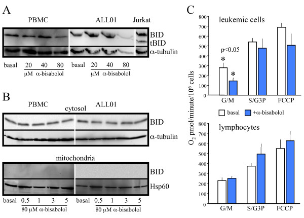
Background: We previously demonstrated that the plant-derived agent α-bisabolol enters cells via lipid rafts, binds to the pro-apoptotic Bcl-2 family protein BID, and may induce apoptosis. Here we studied the activity of α-bisabolol in acute leukemia cells.

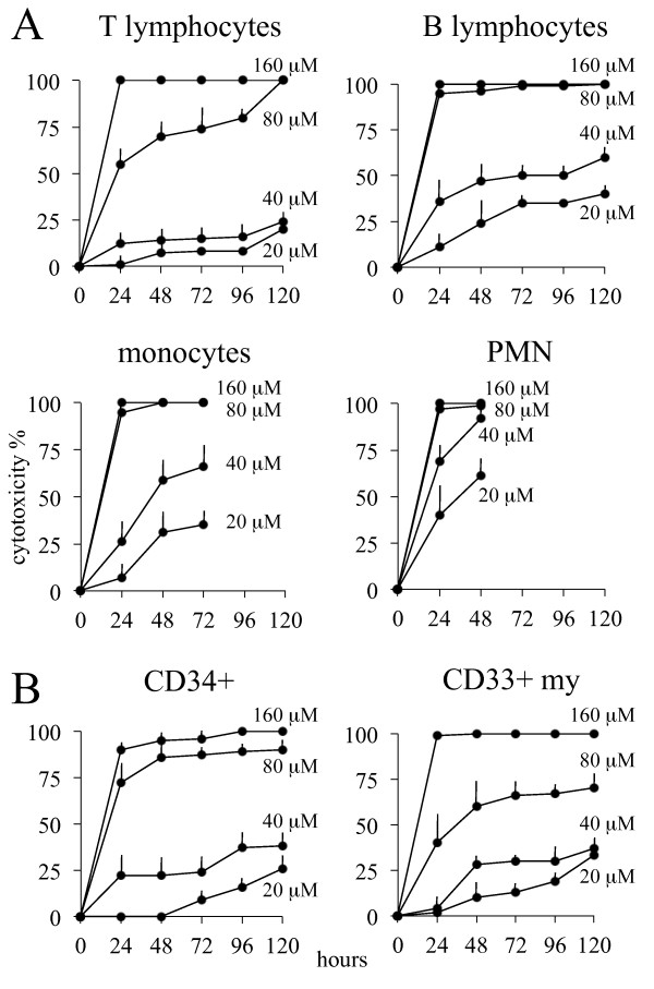
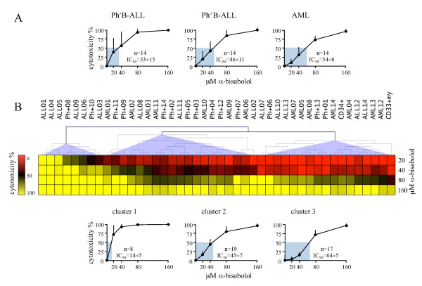
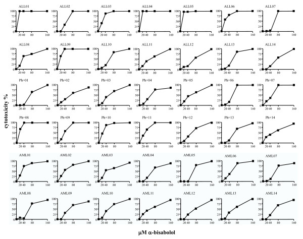
Methods: We tested ex vivo blasts from 42 acute leukemias (14 Philadelphia-negative and 14 Philadelphia-positive B acute lymphoid leukemias, Ph-/Ph+B-ALL; 14 acute myeloid leukemias, AML) for their sensitivity to α-bisabolol in 24-hour dose-response assays. Concentrations and time were chosen based on CD34+, CD33+my and normal peripheral blood cell sensitivity to increasing α-bisabolol concentrations for up to 120 hours.

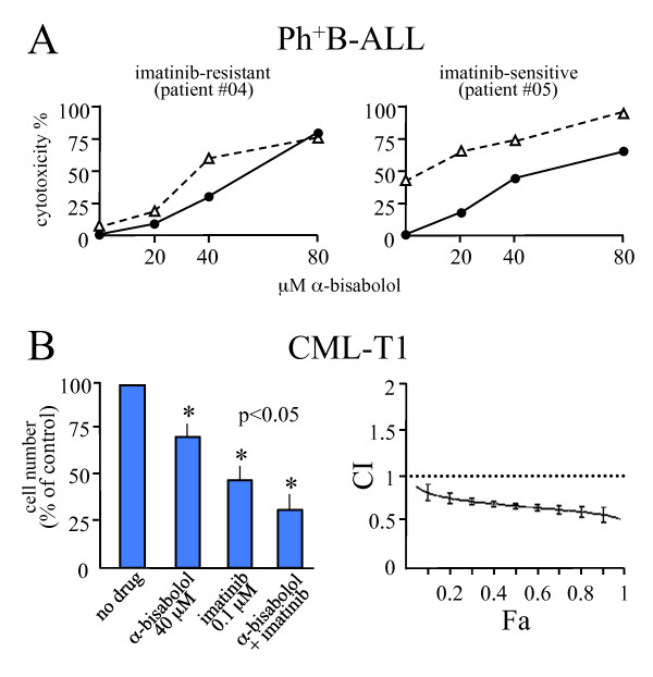
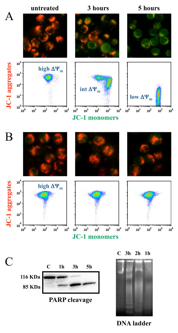
Results: A clustering analysis of the sensitivity over 24 hours identified three clusters. Cluster 1 (14 ± 5 μM α-bisabolol IC50) included mainly Ph-B-ALL cells. AML cells were split into cluster 2 and 3 (45 ± 7 and 65 ± 5 μM IC50). Ph+B-ALL cells were scattered, but mainly grouped into cluster 2. All leukemias, including 3 imatinib-resistant cases, were eventually responsive, but a subset of B-ALL cells was fairly sensitive to low α-bisabolol concentrations. α-bisabolol acted as a pro-apoptotic agent via a direct damage to mitochondrial integrity, which was responsible for the decrease in NADH-supported state 3 respiration and the disruption of the mitochondrial membrane potential.

Conclusion: Our study provides the first evidence that α-bisabolol is a pro-apoptotic agent for primary human acute leukemia cells.


Figures

Figure 1
Figure 1
α-bisabolol structure and solubilization in the culture medium. (A) α-bisabolol is a small oily sesquiterpene alcohol with a molecular mass of 222.37 Da. (B) 250 μM α-bisabolol was added to culture medium: concentration raised during the first 3 hours, then lowered to around 65% of the initially added α-bisabolol after 24 hours. (C) By this time, the linear function relating added to measured concentrations of α-bisabolol shows that the incremental ratio was 0.65 for 14 evaluations representing a double series of 7 scaled concentrations tested by a RP-HPLC method. Each point is the mean ± SD of 2 measurements.
Figure 2
Figure 2
Cytotoxicity of α-bisabolol in normal hematologic cells. (A) Peripheral blood cells. (B) Bone marrow stem cells. Time- and dose-response curves between 20 and 160 μM α -bisabolol in the 120-hour cytotoxicity assays. Means ± SD of 5 normal donors are depicted.
Figure 3
Figure 3
24-hourcytotoxicity of α-bisabolol in primary blasts from 42 acute leukemias. (A) α-bisabolol activity against blasts ex vivo, here grouped by diagnosis. The corresponding IC50 values and number of cases (n) are shown. The differences between the Ph-B-ALL sensitivity curves and the other ones were statistically significant (p < 0.05). Each point is the mean ± SD of 14 cases. (B) α-bisabolol sensitivity clustering analysis. The samples were grouped by complete linkage hierarchical clustering algorithm available in MultiExperiment Viewer http://www.tm4.org/mev/. The heat map was obtained by subtracting spontaneous mortality to scaled α-bisabolol 24-hour cytotoxicity expressed as percentage. Three main groups of patients were identified based on their cytotoxicity response. The Ph-B-ALL (ALL) cases shared the highest sensitivity and were grouped mainly in the first sensitivity cluster, whereas AML cases were split into two groups with intermediate and lower sensitivities. Ph+B-ALL (Ph+) cells were scattered among the three groups, although they were mainly clustered in the second group. At the bottom, the 24-hour dose-response curves of the three sensitivity clusters are depicted, and the corresponding IC50 values and number of cases (n) are shown. The differences between the curves were statistically significant (p < 0.05).
Figure 4
Figure 4
24-hour cytotoxicity of α-bisabolol in each individual case. 14 Ph-B-ALL, 14 Ph+B-ALL, and 14 AML cell samples were treated with 20, 40, 80, and 160 μM α-bisabolol over 24 hours. Captions identify the cases in Table 1 and in Figure 3B (clustering analysis).
Figure 5
Figure 5
24-hour cytotoxicity of α-bisabolol in Ph+ cells as compared to imatinib mesylate. (A) Scaled α-bisabolol alone (solid line) or in combination with 3 μM imatinib mesylate (dashed line) in 2 representative cases out of 10 (Ph+B-ALL #04 and #05 in Table 1, Figure 3B and Figure 4, where α-bisabolol concentrations are represented up to 160 μM). The imatinib mesylate-dependent cytotoxicity is indicated at point 0,0. Cells resistant to imatinib mesylate were sensitive to α-bisabolol. In cells sensitive to imatinib mesylate, α-bisabolol potentiated the effect of the other drug. (B) Analysis of synergism between imatinib and α-bisabolol in the imatinib-sensitive BCR/ABL+ human cell line CML-T1. Left side. Effects of 40 μM α-bisabolol and 0.1 μM imatinib, alone and combined, on proliferation of the cell line. Means ± SD of 5 experiments. Right side. Plot showing the corrisponding combination index (CI) vs. the fraction affected (Fa). CI values are <1, indicating that the two drugs are synergistic. Bars represent the variability of effects according to the sequential deletion analysis [16].
Figure 6
Figure 6
BID and NADH-supported state 3 respiration in normal PBMCs and leukemic blasts treated with α-bisabolol. (A) 24-hour α-bisabolol did not induced the cleavage of BID (full length 22 kDa, cleaved 15 kDa) at any concentration. Etoposide-treated Jurkat cells were used as a positive control for tBID. (B) No BID translocation was detected in mitochondrial fraction at different times and solubilized doses of α-bisabolol. α-tubulin and Hsp60 were used as markers for the cytosol and mitochondria fractions, respectively. A representative case is shown. (C) Permeabilized leukemic cells and healthy lymphocytes were incubated for 10 minutes in respiration buffer at 30°C in the presence or in the absence of 3 μM α-bisabolol. In treated leukemic cells, the G/M oxygen consumption was clearly lower than in untreated leukemic controls (p < 0.05). The S/G3P oxygen consumption was not modified by treatment, and the mitochondrial respiration was not stimulated by FCCP addition. This is in line with a direct effect of α-bisabolol on mitochondrial integrity. Healthy lymphocyte respiration was not affected by treatment. G/M: glutamate plus malate; S/G3P: succinate plus glycerol-3-phosphate; FCCP: carbonylcyanide-4-(trifluoromethoxy)-phenyl-hydrazone. Means ± SD of 6 leukemias and 6 normal donors are depicted.
Figure 7
Figure 7
α-bisabolol-induced mitochondrial damage in primary leukemic blasts. Cells were stained with JC-1. In non-damaged cells, JC-1 forms red-emitting aggregates in the mitochondrial matrix. A loss of red fluorescence and an increase in cytoplasmic green-emitting monomers signal the disruption of the mitochondrial transmembrane potential (ΔΨm). (A) The representative case Ph-B-ALL #01 is shown out of the 6 leukemias tested. Microscopy (magnification, × 400). Whereas untreated leukemic blasts showed well-polarized mitochondria marked by punctated red fluorescent staining, blasts treated with 40 μM α-bisabolol had staining that was quite completely replaced by diffuse green fluorescence, indicating loss of ΔΨm. Flow cytometry. Untreated blasts with well-polarized mitochondria localized in the upper region of the plot (high ΔΨm). Blasts exposed to 40 μM α-bisabolol shifted right and downward (intermediate and low ΔΨm), due to the progressive dislocation of JC-1 from the mitochondria to the cytoplasm, which signaled the disruption of the mitochondrial ΔΨm. (B) Both untreated and α-bisabolol-treated normal lymphocytes used as a negative control maintained well-polarized mitochondria and did not undergo apoptosis. Apoptosis of leukemic blasts was also documented by (C) PARP cleavage and DNA laddering in the same representative case depicted in (A).

Similar articles

Cited by

References

    1. Cavalieri E, Mariotto S, Fabrizi C, De Prati AC, Gottardo R, Leone S, Berra LV, Lauro GM, Ciampa AR, Suzuki H. α-bisabolol, a non-toxic natural compound, strongly induces apoptosis in glioma cells. Biochem Biophys Res Commun. 2004;315:589–594. doi: 10.1016/j.bbrc.2004.01.088. - DOI - PubMed
    1. Costarelli L, Malavolta M, Giacconi R, Cipriano C, Gasparini N, Tesei S, Pierpaoli S, Orlando F, Suzuki H, Perbellini L, Piacenza F, Emanuelli M, Mocchegiani E. In vivo effect of alpha-bisabolol, a nontoxic sesquiterpene alcohol, on the induction of spontaneous mammary tumors in HER-2/neu transgenic mice. Oncol Res. 2010;18:409–418. doi: 10.3727/096504010X12671222663557. - DOI - PubMed
    1. Darra E, Abdel-Azeim S, Manara A, Shoji K, Marechal JD, Mariotto S, Cavalieri E, Perbellini L, Pizza C, Perahia D, Crimi M, Suzuki H. Insight into the apoptosis-inducing action of α-bisabolol towards malignant tumor cells: Involvement of lipid rafts and Bid. Arch Biochem Biophys. 2008;476:113–123. doi: 10.1016/j.abb.2008.02.004. - DOI - PubMed
    1. Tallman MS, Gilliland DG, Rowe JM. Drug therapy for acute myeloid leukemia. Blood. 2005;106:1154–1163. doi: 10.1182/blood-2005-01-0178. - DOI - PubMed
    1. Appelbaum FR, Rosenblum D, Arceci RJ, Carroll WL, Breitfeld PP, Forman SJ, Larson RA, Lee SJ, Murphy SB, O'Brien S, Radich J, Scher NS, Smith FO, Stone RM, Tallman MS. End points to establish the efficacy of new agents in the treatment of acute leukemia. Blood. 2007;109:1810–1816. doi: 10.1182/blood-2006-08-041152. - DOI - PubMed

Publication types

MeSH terms