Review
. 2011 May;30(4):235-42.
doi: 10.1016/j.matbio.2011.03.005. Epub 2011 Apr 13.

Early growth response transcription factors: key mediators of fibrosis and novel targets for anti-fibrotic therapy

Affiliations
Review

Early growth response transcription factors: key mediators of fibrosis and novel targets for anti-fibrotic therapy

Swati Bhattacharyya et al. Matrix Biol. 2011 May.

Abstract

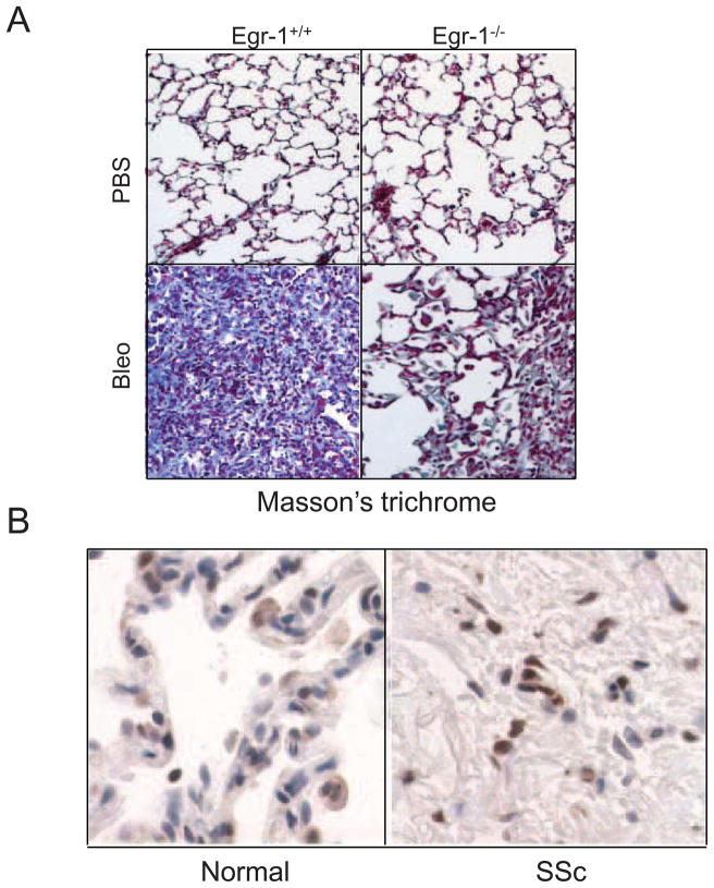
Fibrosis is a deregulated and ultimately defective form of tissue repair that underlies a large number of chronic human diseases, as well as obesity and aging. The pathogenesis of fibrosis involves multiple cell types and extracellular signals, of which transforming growth factor-ß (TGF-ß) is pre-eminent. The prevalence of fibrosis is rising worldwide, and to date no agents has shown clinical efficacy in the attenuating or reversing the process. Recent studies implicate the immediate-early response transcription factor Egr-1 in the pathogenesis of fibrosis. Egr-1 couples acute changes in the cellular environment to sustained alterations in gene expression, and mediates a broad spectrum of biological responses to injury and stress. In contrast to other ligand-activated transcription factors such as NF-κB, c-jun and Smad2/3 that undergo post-translational modification such as phosphorylation and nuclear translocation, Egr-1 activity is regulated via its biosynthesis. Aberrant Egr-1 expression or activity is implicated in cancer, inflammation, atherosclerosis, and ischemic injury and recent studies now indicate an important role for Egr-1 in TGF-ß-dependent profibrotic responses. Fibrosis in various animal models and human diseases such as scleroderma (SSc) and idiopathic pulmonary fibrosis (IPF) is accompanied by aberrant Egr-1 expression. Moreover Egr-1 appears to be required for physiologic and pathological connective tissue remodeling, and Egr-1-null mice are protected from fibrosis. As a novel profibrotic mediator, Egr-1 thus appears to be a promising potential target for the development of anti-fibrotic therapies.


Figures

Fig. 1
Fig. 1
Egr-1 and related proteins of the Egr gene family. Egr-1 consists of activation domains, an inhibitory (Nab Interaction) domain and a DNA-binding domain.
Fig. 2
Fig. 2
Regulation of TGF-β responses by Egr-1 and its inhibitor. A. TGF-β binds to the TβR1–TβR2 complex causing activation of Smad2/3. The pSmad2/3-Smad4 complex then translocates into the nucleus, where it binds to Smad binding elements (SBE) of target genes. The coactivator p300 is recruited to the Smad complex to activate COL1A2 transcription. TGF-β also activates a non-Smad pathway via c-Abl which in turn stimulates Egr-1 via MAP kinase. Egr-1 then binds to the Egr-1 binding element (EBS) to further stimulate COL1A2 transcription. B: Nab2 modulates Egr-1 activity and TGF-β signaling. In unstimulated fibroblasts (basal state), small amounts of Egr-1 and Nab2 are constitutively associated with the COL1A2 promoter (left upper panel). During early TGF-β stimulation (30min–4h) (right upper panel), Egr-1 is induced and Egr-1 is recruited to the COL1A2 promoter, where it enhances histone H4 hyperacetylation and stimulates transcription. Sustained TGF-β stimulation (2–48h) (left lower panel) leads to increased Nab2 expression and its accumulation in the Egr-1-COL1A2 transcriptional complex, where recruitment of HDAC1 resulting in H4 histone deacetylation and transcriptional silencing. In pathological fibrosis associated with constitutive Egr-1 expression (exemplified by scleroderma fibroblasts), defective Nab2 induction or function might results in unopposed Egr-1 signaling and target gene transcription (right lower panel).
Fig. 2
Fig. 2
Regulation of TGF-β responses by Egr-1 and its inhibitor. A. TGF-β binds to the TβR1–TβR2 complex causing activation of Smad2/3. The pSmad2/3-Smad4 complex then translocates into the nucleus, where it binds to Smad binding elements (SBE) of target genes. The coactivator p300 is recruited to the Smad complex to activate COL1A2 transcription. TGF-β also activates a non-Smad pathway via c-Abl which in turn stimulates Egr-1 via MAP kinase. Egr-1 then binds to the Egr-1 binding element (EBS) to further stimulate COL1A2 transcription. B: Nab2 modulates Egr-1 activity and TGF-β signaling. In unstimulated fibroblasts (basal state), small amounts of Egr-1 and Nab2 are constitutively associated with the COL1A2 promoter (left upper panel). During early TGF-β stimulation (30min–4h) (right upper panel), Egr-1 is induced and Egr-1 is recruited to the COL1A2 promoter, where it enhances histone H4 hyperacetylation and stimulates transcription. Sustained TGF-β stimulation (2–48h) (left lower panel) leads to increased Nab2 expression and its accumulation in the Egr-1-COL1A2 transcriptional complex, where recruitment of HDAC1 resulting in H4 histone deacetylation and transcriptional silencing. In pathological fibrosis associated with constitutive Egr-1 expression (exemplified by scleroderma fibroblasts), defective Nab2 induction or function might results in unopposed Egr-1 signaling and target gene transcription (right lower panel).
Fig. 3
Fig. 3
Normal human dermal fibroblasts were infected with Ad-EGFP or Ad-Egr-1m (100 MOI). At the end of 24 or 48 h incubation, total RNA was isolated and subjected to genomewide transcriptional analysis using Illumina Microarray chips. Genome-wide expression profiling of Egr-1 regulated genes. GO analysis showing biological processes significantly enriched with Egr-1 regulated genes(p < 0.001). A subset of the biological processes is shown.
Fig. 4
Fig. 4
Egr-1 modulates lung fibrosis in mice and human. A. Reduced lung fibrosis in mice lacking Egr-1. Egr-null mice and wild-type littermates received daily s.c. injections of bleomycin or PBS for 14 days. Fourteen days after the last injections, the lungs were harvested and examined with Masson’s trichrome stain (Original magnification x400). B. Elevated Egr-1 expression in SSc lung. Lesional lung tissue from patients with SSc- associated end-stage fibrosis (n=3) and healthy controls (n=3) were examined by immunohistochemistry with antibodies to Egr-1. Reprentative images are shown (original magnification x1000).

References

    1. Baron V, Adamson ED, Calogero A, Ragona G, Mercola D. The transcription factor Egr1 is a direct regulator of multiple tumor suppressors including TGFbeta1, PTEN, p53, and fibronectin. Cancer Gene Ther. 2006;13:115–124. - PMC - PubMed
    1. Bea F, Blessing E, Shelley MI, Shultz JM, Rosenfeld ME. Simvastatin inhibits expression of tissue factor in advanced atherosclerotic lesions of apolipoprotein E deficient mice independently of lipid lowering: potential role of simvastatin-mediated inhibition of Egr-1 expression and activation. Atherosclerosis. 2003;167:187–194. - PubMed
    1. Beattie J, Allan GJ, Lochrie JD, Flint DJ. Insulin-like growth factor-binding protein-5 (IGFBP-5): a critical member of the IGF axis. Biochem J. 2006;395:1–19. - PMC - PubMed
    1. Bhattacharyya S, Chen SJ, Wu M, Blankenship MW, Ning H, Lakos G, Mori Y, Chang E, Nihijima C, Takehara K, Feghali-Bostwick C, Varga J. Smad-Independent Transforming Growth Factor-β Regulation of Early Growth Response-1 and Sustained Expression in Fibrosis. Implications for Scleroderma. Am J Path. 2008;173:1085–1099. - PMC - PubMed
    1. Bhattacharyya S, Ishida W, Wu M, Wilkes M, Mori Y, Hinchcliff M, Leof E, Varga J. A non-Smad mechanism of fibroblast activation by transforming growth factor-b via c-Abl and Egr-1: selective modulation by imatinib mesylate. Oncogene. 2008:1–13. - PMC - PubMed

MeSH terms

Substances