. 2011 Apr 22;34(4):566-78.
doi: 10.1016/j.immuni.2011.03.018.

Interleukin-10 signaling in regulatory T cells is required for suppression of Th17 cell-mediated inflammation

Affiliations

Interleukin-10 signaling in regulatory T cells is required for suppression of Th17 cell-mediated inflammation

Ashutosh Chaudhry et al. Immunity. .

Abstract

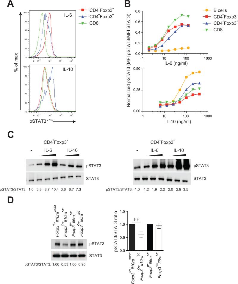
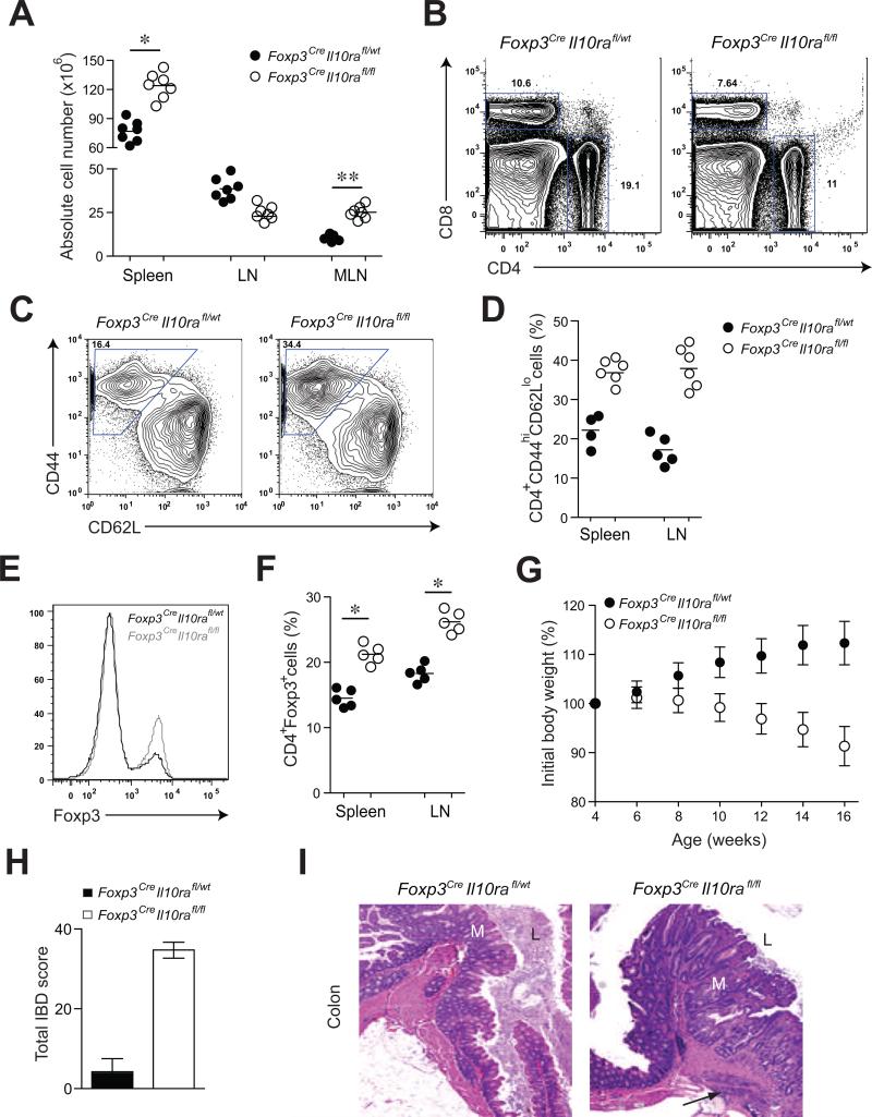
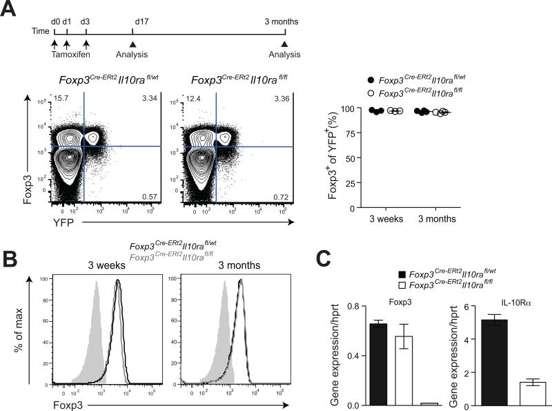
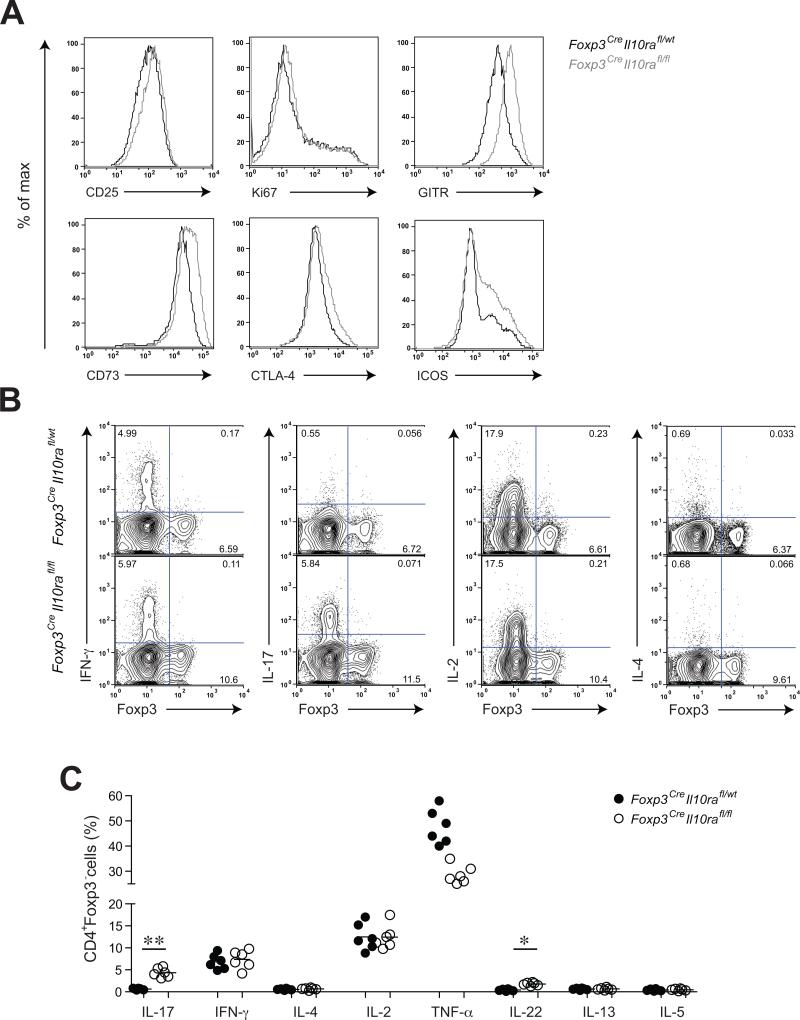
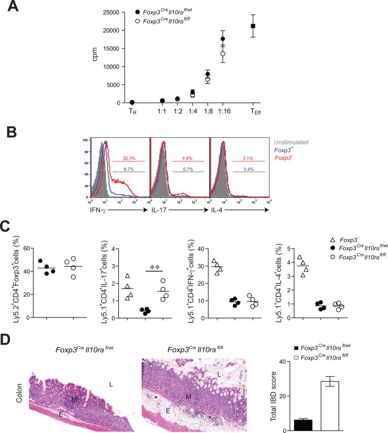
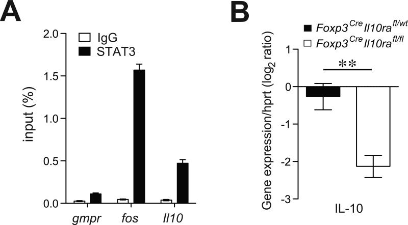
Effector CD4+ T cell subsets, whose differentiation is facilitated by distinct cytokine cues, amplify the corresponding type of inflammatory response. Regulatory T (Treg) cells integrate environmental cues to suppress particular types of inflammation. In this regard, STAT3, a transcription factor essential for T helper 17 (Th17) cell differentiation, is necessary for Treg cell-mediated control of Th17 cell responses. Here, we showed that anti-inflammatory interleukin-10 (IL-10), and not proinflammatory IL-6 and IL-23 cytokine signaling, endowed Treg cells with the ability to suppress pathogenic Th17 cell responses. Ablation of the IL-10 receptor in Treg cells resulted in selective dysregulation of Th17 cell responses and colitis similar to that observed in mice harboring STAT3-deficient Treg cells. Thus, Treg cells limit Th17 cell inflammation by serving as principal amplifiers of negative regulatory circuits operating in immune effector cells.


Figures

Figure 1
Figure 1. IL-10 induces robust STAT3 phosphorylation in Treg cells
(A) Flow cytometric analysis of pSTAT3 (Y705) amounts after treatment of splenic CD4+Foxp3- T cells, CD4+Foxp3+ Treg cells and CD8 T cells with recombinant IL-6 (16 ng/ml) or IL-10 (100 ng/ml) for 15 mins. Analysis of pSTAT3 levels by either flow cytometry (B) or western blotting (C) after stimulation of indicated cells with titrating doses (3, 10 and 15 ng/ml for IL-6; 50, 150 and 300 ng/ml for IL-10) of recombinant IL-6 or IL-10. (D) Immunoblot detection of pSTAT3 and STAT3 in Treg cells isolated from IL-10R or IL-6R deficient and sufficient mice. Data are representative of four independent experiments.
Figure 2
Figure 2. Treg cell-specific IL-10R deletion results in severe intestinal inflammation
(A, B) Spleen and lymph node cellularity in Foxp3CreIl10rafl/wt and Foxp3CreIl10rafl/fl mice (** p<0.001; * p<0.01). (C, D) Frequencies of activated T cells (E, F) Absolute numbers of CD4+Foxp3+ T cells and (G) body weights of Foxp3CreIl10rafl/fl mice (n=7). (H) IBD scores derived from evaluation of colon and cecum from 14-16 week-old Foxp3CreIl10rafl/wt and Foxp3CreIl10rafl/fl mice (n=4/group). (I) Representative H&E stained sections of large bowel at the level of the ileocecal-colic junction from Foxp3CreIl10rafl/wt and Foxp3CreIl10rafl/fl mice. Note: Mucosa (M) with expanded irregular, hyperplastic crypts, glandular loss and fibrosis with multifocal transmural inflammation (arrow). Lumen (L). Original magnification, 5x (n=7).
Figure 3
Figure 3. IL-10R ablation does not affect Foxp3 expression and Treg cell lineage stability
(A) Flow cytometric analysis of Foxp3 and YFP expression on splenic CD4+ T cells in Foxp3CreIl10rafl/flR26Y and control littermate mice. (B) Expression of Foxp3 in flow cytometry-sorted CD4+YFP+ T cells from indicated mice. (C) Quantitative PCR (qPCR) analysis of Foxp3 and IL-10Rα expression in flow cytometry-sorted CD4+YFP+ T cells. The grey bar represents CD4+YFP- T cells. The results represent the mean+/-SD of relative expression values for the indicated genes relative to hypoxanthine-guanine phosphoribosyl transferase. Data are representative of three independent experiments.
Figure 4
Figure 4. Increased Treg cell activation and selective dysregulation of Th17 cell responses in Foxp3CreIl10rafl/fl mice
(A) Flow cytometric analysis of Treg cell activation markers and putative effector molecules on splenic CD4+Foxp3+ T cells in Foxp3CreIl10rafl/fl and control littermate mice. (B, C) Flow cytometric analysis of cytokine production by splenic CD4+Foxp3- T cells in Foxp3CreIl10rafl/wt and Foxp3CreIl10rafl/fl mice after PMA and Ionomycin treatment for 4-6 hours (** p<0.001; * p<0.01).
Figure 5
Figure 5. IL-10R-deficient Treg cells efficiently control in vitro T cell proliferation but fail to suppress Th17 cell differentiation and colitis
(A) IL-10R-deficient and -sufficient Treg cell-mediated suppression of effector T cell (TEff) proliferative responses in vitro. (B) Frequencies of Th1, Th2 and Th17 skewed cells among the transferred T cell population isolated from the Foxp3- mice. (C) Frequencies of splenic Treg cells, CD4+IL-17+Foxp3- T cells, CD4+IFN-γ+ Foxp3- T cells, and CD4+IL-4+Foxp3- T cells within the indicated donor-derived population 5-6 weeks after co-transfer of either Foxp3CreIl10rafl/wt or Foxp3CreIl10rafl/fl Treg cells with Foxp3- CD4+ T cells into Rag2-/- recipients. (D) Representative H&E stained sections of colon collected from recipient mice 5-6 week after adoptive T cell transfer (original magnification, 10x). Note: Mucosa (M), submucosal edema (E), dilated lymphatics (*) and lumen (L).
Figure 6
Figure 6. IL-10R signaling is required for IL-10 expression by Treg cells
(A) qPCR analysis of STAT3-bound chromatin isolated from wild type Treg cells using primers corresponding to the indicated genes. House-keeping gene Gmpr was used as a specificity control while IgG-immunoprecipitated chromatin was used as a negative control. (B) qPCR analysis of relative expression of indicated genes in Treg cells isolated from either Foxp3CreIl10rafl/wt or Foxp3CreIl10rafl/fl mice. The results represent the mean+/-SD of relative expression values for the indicated genes relative to hypoxanthineguanine phosphoribosyl transferase.

Comment in

References

    1. Adamson AS, Collins K, Laurence A, O'Shea JJ. The Current STATus of lymphocyte signaling: new roles for old players. Curr Opin Immunol. 2009;21:161–166. - PMC - PubMed
    1. Ahern PP, Izcue A, Maloy KJ, Powrie F. The interleukin-23 axis in intestinal inflammation. Immunol Rev. 2008;226:147–159. - PubMed
    1. Brand S. Crohn's disease: Th1, Th17 or both? The change of a paradigm: new immunological and genetic insights implicate Th17 cells in the pathogenesis of Crohn's disease. Gut. 2009;58:1152–1167. - PubMed
    1. Burich A, Hershberg R, Waggie K, Zeng W, Brabb T, Westrich G, Viney JL, Maggio-Price L. Helicobacter-induced inflammatory bowel disease in IL-10- and T cell-deficient mice. Am J Physiol Gastrointest Liver Physiol. 2001;281:G764–778. - PubMed
    1. Chaudhry A, Rudra D, Treuting P, Samstein RM, Liang Y, Kas A, Rudensky AY. CD4+ regulatory T cells control TH17 responses in a Stat3-dependent manner. Science. 2009;326:986–991. - PMC - PubMed

Publication types