Review
. 2011 Jul;156(2-3):103-14.
doi: 10.1016/j.bpc.2011.03.010. Epub 2011 Apr 5.

Metalloregulatory proteins: metal selectivity and allosteric switching

Affiliations
Review

Metalloregulatory proteins: metal selectivity and allosteric switching

Hermes Reyes-Caballero et al. Biophys Chem. 2011 Jul.

Abstract

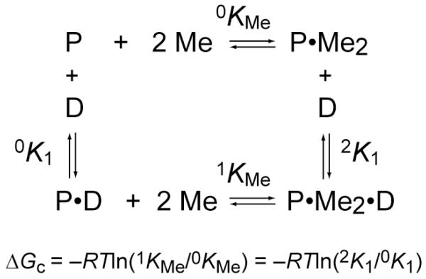
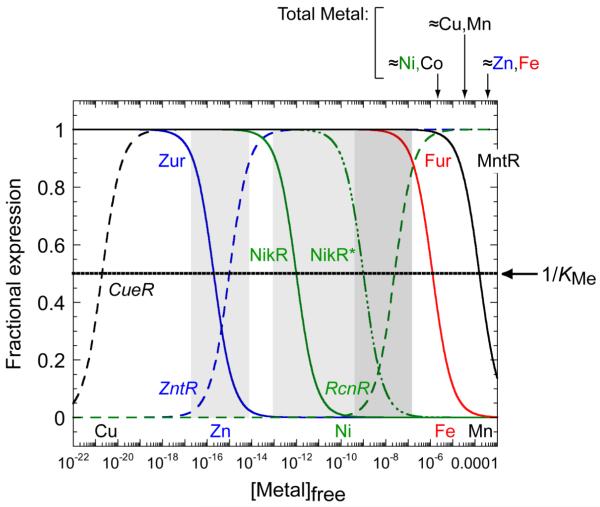
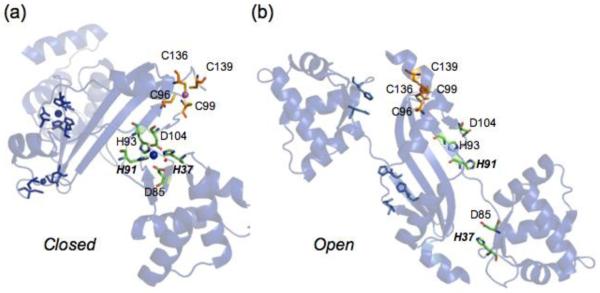
Prokaryotic organisms have evolved the capacity to quickly adapt to a changing and challenging microenvironment in which the availability of both biologically required and non-essential transition metal ions can vary dramatically. In all bacteria, a panel of metalloregulatory proteins controls the expression of genes encoding membrane transporters and metal trafficking proteins that collectively manage metal homeostasis and resistance. These "metal sensors" are specialized allosteric proteins, in which the direct binding of a specific or small number of "cognate" metal ion(s) drives a conformational change in the regulator that allosterically activates or inhibits operator DNA binding, or alternatively, distorts the promoter structure thereby converting a poor promoter to a strong one. In this review, we discuss our current understanding of the features that control metal specificity of the allosteric response in these systems, and the role that structure, thermodynamics and conformational dynamics play in mediating allosteric activation or inhibition of DNA binding.


Figures

Figure 1
Figure 1
A coupled equilibrium thermodynamic scheme that defines the relationship of all four allosteric states of the homodimer repressor (P) in equilibrium with DNA operator (D) and metal ions (Me), with a limiting stoichiometry of two metals per dimer. The allosteric coupling free energy, ΔGc, is defined as indicated.
Figure 2
Figure 2
A graphical representation of the hypothesis that metal affinity for individual or pairs of metal sensor proteins (1/KMe) defines the ability of the cytoplasm to buffer biologically required transitions metal ions. Cu(I), black; Zn(II), blue; Ni(II), green; Fe(II), red; Mn(II), black. See Table 1 for KMe values for the regulators indicated. Efflux regulators are italicized, with transcriptional response curves (derepression or activation) represented by the dashed lines; uptake repressors are in straight font and transcriptional response curves (corepression) represented by the solid lines. NikR* represents filling of the secondary, low affinity sites which give rise to full repression of the high affinity nickel uptake system [48]. Approximate total cell-associated concentrations measured in E. coli under aerobic growth conditions on a minimal media are shown above the figure [28]; recent studies suggest that these concentrations are ≈5-fold larger in S. pneumoniae, which also exhibits a high Mn(II) quota relative to Fe(II) [24]. Note that this model is predicated on the simple notion that metal sensors are capable of “scanning” the cytoplasm for metal; this may not be the case [29, 139].
Figure 3
Figure 3
Native metal coordination geometries for selected bacterial metalloregulatory proteins. A colored sphere represent the metal ion unless is otherwise indicated. (a) E. coli CueR, Cu(I) linear (PDB ID:1Q05), (b) M. tuberculosis CsoR, trigonal Cu(I) (PDB ID: 2HH7), (c) B. subtilis MntR, penta-coordinate dinuclear Mn(II), H2O molecules are represented as cyan spheres (PDB ID: 1ON1), (d) E. coli ZntR, tetrahedral dinuclear Zn(II) (PDB ID: 1Q08), (e) E. coli NikR, square planar Ni(II) (PDB ID: 2HZV), (f) S. elongatus PC7942 SmtB, Zn(II) tetrahedral (PDB ID: 1R22). Graphical representation was created using PyMol [140].
Figure 4
Figure 4
Illustration of a potential hydrogen bonding pathway in CzrA that links the metal binding and DNA binding sites. (a) Ribbon representation of the solution structure of CzrA bound to DNA with key residues highlighted [89] The blue arrow defines the distance (≈10 Å) between the L68′ and H97 on opposite protomers that are proposed to hydrogen bond in the allosterically inhibited Zn2-bound state (see panel b). (b) Two hydrogen bonds are proposed to link the NHε2 of His97 with the main chain C=O of L63′ in the DNA binding αR helix [91]. (c) 15N-1H correlation for the HNε2 group of H97 observed in a simple 15N-1H HSQC spectrum of Zn2-CzrA, indicative of slow exchange with solvent in the allosterically inhibited metal-bound state, consistent with a hydrogen bonding interaction. This correlation is found in all first coordination shell mutants of CzrA that are capable of negatively regulating DNA binding upon Zn(II) binding [63], and is lost in apo-CzrA [91].
Figure 5
Figure 5
The first coordination shell of a liganding histidine as an allosteric trigger in moving from the (a) “closed” PerRMnZn (3F89) and (b) “open” apo-Zn (2FE3) structures. The structure shows two metal binding sites occupied by Mn(II) (blue) and Zn(II) (magenta). Mn(II) binds to the regulatory site in a distorted square pyramidal geometry involving ligands H37 and H91 and in the Fe(II) complex, an open coordination site that accommodate H2O2. Zn(II) binds to a thiolate rich site that plays no role in the catalysis [95, 96], but is essential for structural integrity of the dimer [99].
Figure 6
Figure 6
Global energetics of the step-wise coupled equilibrium that describes the allosteric effect of Zn(II) binding to CzrA free in solution and when bound to DNA. Thermodynamic parameters determined by isothermal titration calorimetry (ITC) are shown [74] at pH 7.0, 0.4 M NaCl. 25.0 °C.. This is an expanded thermodynamic linkage scheme relative to that outlined in Fig. 1. CzrO, czr DNA operator. K1, K2, K5 and K6 are KZni from each step in the equilbrium where ΔGi = −RTlnKZni. Allosteric (heterotropic) coupling free energies, ΔGci are obtained as follows: ΔGc1 = ΔG5 − ΔG1 (defined by the yellow box); ΔGc2 = ΔG6 − ΔG2 (defined by the green box); ΔGct = (ΔG5 + ΔG6) − (ΔG1 + ΔG2) = ΔGc1 + ΔGc2 = ΔG4 − ΔG3 (overall linkage). See text for other details.
Figure 7
Figure 7
Zinc binds to apo-CzrA homodimer and quenches the conformational dynamics in the core α5, α1 helical regions but also in more peripheral DNA-binding αR helices of the molecule [89]. Spacefilling models of Zn2 CzrA are shown (left, α5 helices on top; right, view from the DNA-binding interface) with residues shaded according to a specific change in dynamics relative to apo-CzrA. Blue, residues for which the order parameter S2 increases (≥0.02); purple, residues for which significant chemical exchange broadening (Rex) is lost on Zn(II) binding; beige, residues for which Rex becomes measurable upon Zn(II) binding; yellow, residues for which S2 decreases on Zn(II) binding. This picture is consistent with significant quenching of the conformational dynamics (indicated by blue, purple residues) far from the allosteric zinc binding sites, relative to those residues which increase their motional disorder (beige, yellow); these may define “hinge” regions in the allosterically inhibited Zn(II) conformational ensemble [90].

References

    1. Berg JM, Godwin HA. Lessons from zinc-binding peptides. Annu Rev Biophys Biomol Struct. 1997;26:357–371. - PubMed
    1. Seneque O, Bonnet E, Joumas FL, Latour JM. Cooperative Metal Binding and Helical Folding in Model Peptides of Treble-Clef Zinc Fingers. Chemistry. 2009;15:4798–4810. - PubMed
    1. Auld DS. Zinc coordination sphere in biochemical zinc sites. Biometals. 2001;14:271–313. - PubMed
    1. Perry JJP, Shin DS, Getzoff ED, Tainer JA. The structural biochemistry of the superoxide dismutases. Biochim Biophys Acta. 2010;1804:245–262. - PMC - PubMed
    1. Ermler U, Grabarse W, Shima S, Goubeaud M, Thauer RK. Active sites of transition-metal enzymes with a focus on nickel. Curr Opin Struct Biol. 1998;8:749–758. - PubMed

Publication types

LinkOut - more resources