

Review
doi: 10.1259/bjr/66754762.
Assessment of post-radiotherapy salivary glands
Affiliations
- PMID: 21511748
- PMCID: PMC3473647
- DOI: 10.1259/bjr/66754762
Item in Clipboard
Review
Assessment of post-radiotherapy salivary glands
Br J Radiol.
2011 May.
Abstract
Salivary glands are usually irradiated during radiotherapy for head and neck cancers, which can lead to radiation-induced damage. Radiation-induced xerostomia (oral dryness) is the most common post-radiotherapy complication for head and neck cancer patients and can reduce the patient's quality of life. Accurate and efficient salivary gland assessment methods provide a better understanding of the cause and degree of xerostomia, and may help in patient management. At present, there are different methods for the assessment of salivary gland hypofunction; however, none of them are considered to be standard procedure. This article reviews the value of common methods in the assessment of post-radiotherapy salivary glands.
Figures
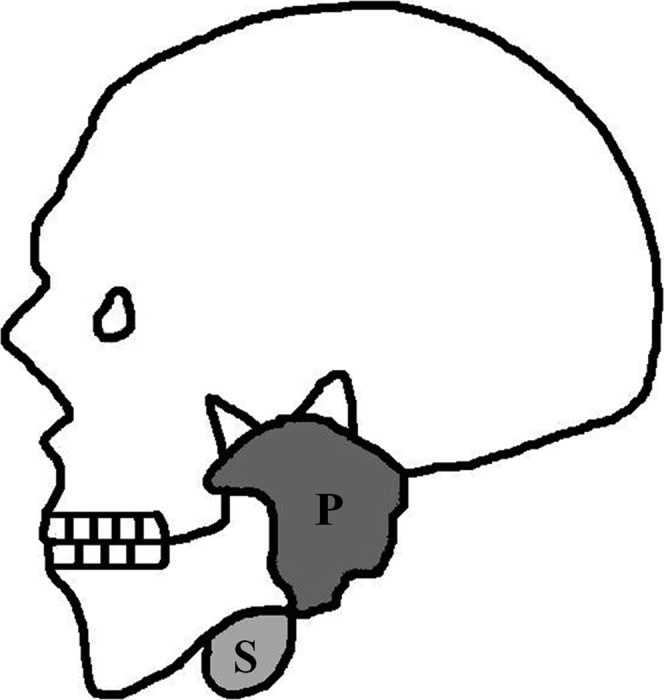
Schematic diagram shows the relative position of parotid gland (P) and submandibular gland (S) in the head and neck region. The parotid gland is located in the retromandibular fossa while the submandibular gland is located under the mandible and in the floor of the oral cavity.
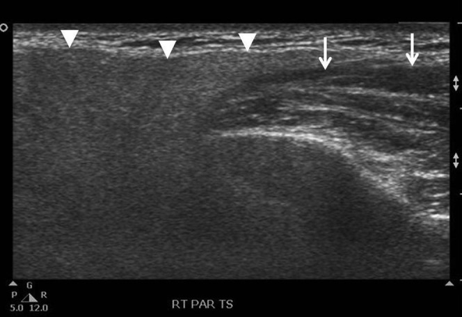
Sonogram shows a transverse scan of a normal parotid gland (arrowheads), which is hyperechoic compared with the adjacent masseter muscle (arrows).
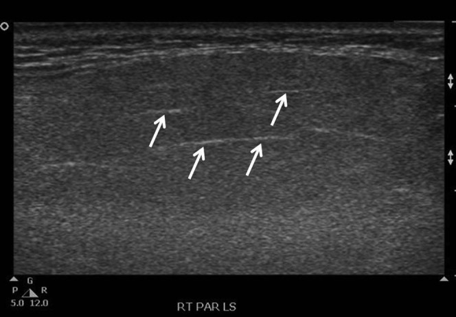
Sonogram shows a longitudinal scan of a normal parotid gland with homogeneous echotexture. The intraglandular ducts (arrows) are marginally seen.
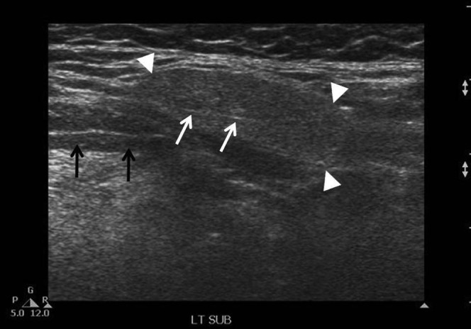
Sonogram shows a transverse scan of a normal submandibular gland (arrowheads) with homogeneous echotexture. The gland is hyperechoic compared with the adjacent mylohyoid muscle (black arrows) and the intraglandular ducts (white arrows) are marginally seen.
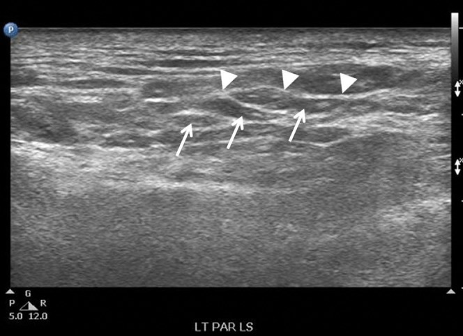
Sonogram shows a longitudinal scan of a parotid gland in a patient treated with conventional radiotherapy. There are multiple hypoechoic areas (arrows) within the gland, and the intraglandular ducts (arrowheads) are obviously seen.

(a) Axial CT scan of skull in a nasopharyngeal carcinoma patient before radiotherapy. The image density of the parotid gland (P) is about the same as the adjacent masseter muscle (M). (b) Axial CT scan of a skull in the same patient after radiotherapy. There is an increased image density of the parotid gland (P) when compared with the pre-radiotherapy scan. The image density of the parotid gland is greater than that of the adjacent masseter muscle (M).
Comment in
-
Post-radiogenic density changes on CT of the salivary gland are time-dependent.Br J Radiol. 2011 Dec;84(1008):1156; author reply 1157. doi: 10.1259/bjr/50052857. Br J Radiol. 2011. PMID: 22101584 Free PMC article. No abstract available.
References
-
- Astreinidou E, Roesink JM, Raaijmakers CPJ, Bartels LW, Witkamp TD, Lagendijk JJ, et al. 3D MR sialography as a tool to investigate radiation-induced xerostomia: feasibility study. Int J Radiat Oncol Biol Phys 2007;68:1310–19 - PubMed
-
- Marmiroli L, Salvi G, Caiazza A, Di Rienzo L, Massaccesi M, Murino P, et al. Dose and volume impact on radiation-induced xerostomia. Rays 2005;30:145–8 - PubMed
-
- Nishimura Y, Nakamatsu K, Shibata T, Kanamori S, Koike R, Okumura M, et al. Importance of the initial volume of parotid glands in xerostomia for patients with head and neck cancers treated with IMRT. Jpn J Clin Oncol 2005;35:375–9 - PubMed
-
- Eisbruch A, Ship JA, Dawson LA, Kim HM, Bradford CR, Terrell JE, et al. Salivary gland sparing and improved target irradiation by conformal and intensity modulated irradiation of head and neck cancer. World J Surg 2003;27:832–7 - PubMed
-
- Coppes RP, Vissink A, Konings AWT. Comparison of radiosensitivity of rat parotid and submandibular glands after different radiation schedules. Radiother Oncol 2002;63:321–8 - PubMed
Publication types
MeSH terms
LinkOut - more resources
Full Text Sources
Medical
